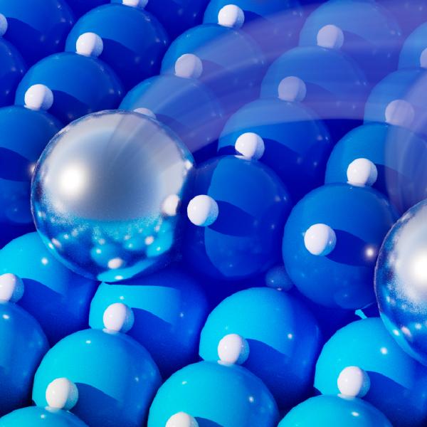
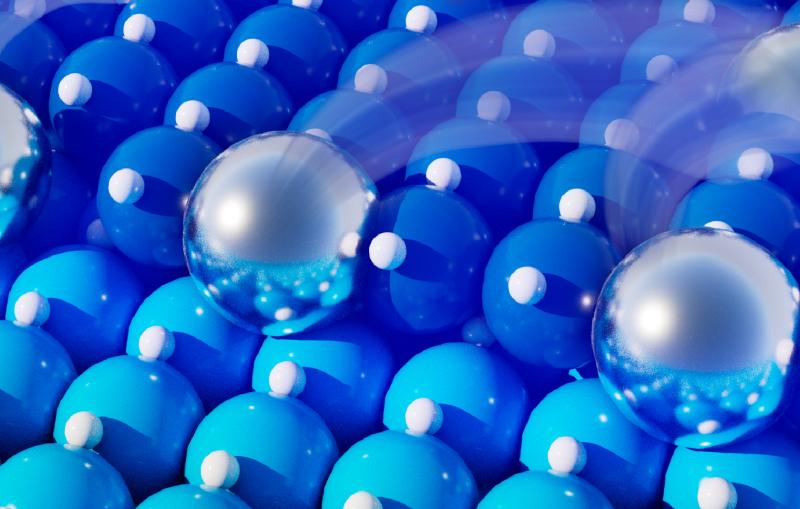
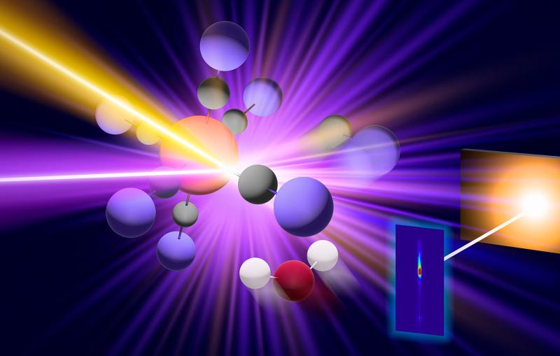
SLAC topics

X-ray light sources and electron imaging RSS feed

X-ray light sources and electron imaging are advanced techniques used to study the structure and properties of materials. X-ray light sources use high-energy photons to produce X-rays, while electron imaging uses high-energy electrons to produce detailed images of samples. 

Browse tagged content

Aerial view of SLAC
Feature

The results should further our understanding of similar reactions with vital roles in chemistry, such as the production of vitamin D in our bodies.

UED transition state
Feature

Chemical reactions often involve intermediate steps that are too fast and complex for us to see  – even using our most advanced scientific instruments...

This is a graphic representation of an intermediate chemical reaction. The image shows the chemical reaction, a laser, X-rays and a detector system.
News Release

After decades of effort, scientists have finally seen the process by which nature creates the oxygen we breathe using SLAC’s X-ray laser.

Photosystem II
Animation

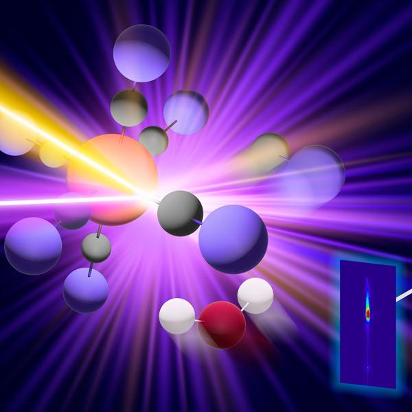
In photosystem II, the water-splitting center cycles through four stable states
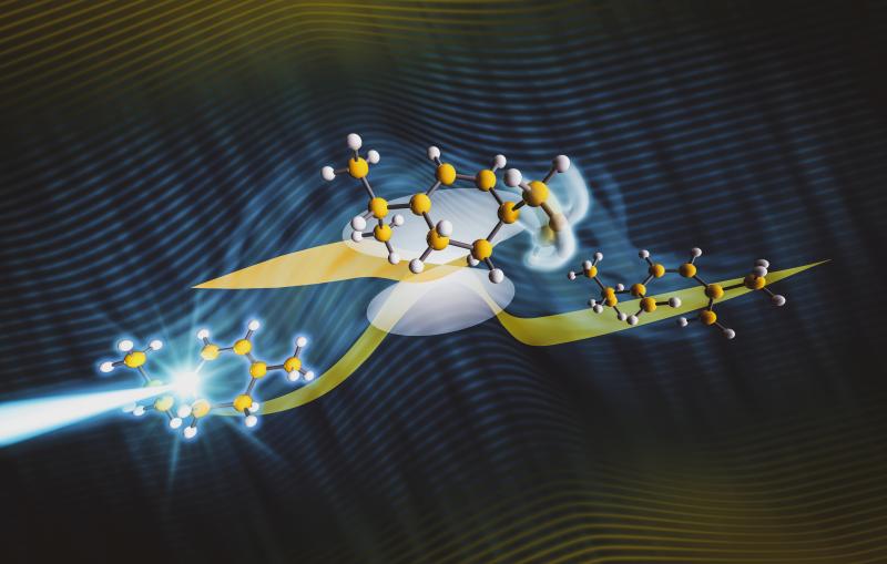
Photosystem II baseball
Feature

A molecule with hooks that can grip and disable the virus’s pesky protease shows potential for fighting infection.

This graphic illustration shows how a SARS-CoV-2 protease attaches to a new molecule. The new molecule is meant to slow the virus inside an infected person.
Feature

This ‘beautiful’ herringbone-like pattern could give rise to unique features that scientists are just starting to explore.

An illustration of a dramatic, herringbone-like pattern in the atomic lattice of a newly created quantum material. Against a black background, calcium atoms are seen as light blue spheres, cobalt atoms in dark blue and oxygen atoms in red. Lines connecting the oxygen atoms represent the atomic lattice.
Illustration

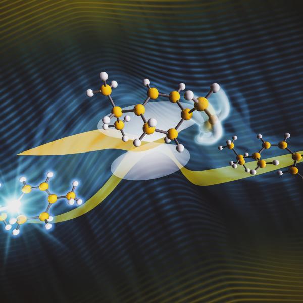
This illustration depicts a herringbone-like pattern in the atomic lattice of a quantum material created by researchers at SLAC and Stanford.

An illustration of a dramatic, herringbone-like pattern in the atomic lattice of a newly created quantum material. Against a black background, calcium atoms are seen as light blue spheres, cobalt atoms in dark blue and oxygen atoms in red. Lines connecting the oxygen atoms represent the atomic lattice.
Feature

Experiments visualize how 2D perovskite structures change when excited.

MeV-UED
Feature

Researchers discover that electrons play a surprising role in heat transfer between layers of semiconductors, with implications for next-generation electronic devices.  

UED electronic bridge
Illustration

Illustration of electrons between layers of 2D semiconductor materials, facilitating rapid heat transfer.

UED electronic bridge
News Release

Researchers used cryo-EM (left) to discover how a chamber in human cells (right) directs protein folding. 

A pom-pom like object with curly tangles in purple and blue shades and yellow tangles at center, reminiscent of a zinnia blossom.
Feature

Once built, the system could produce fast X-ray pulses ten times more powerful than ever before.

illustration of an electron beam traveling through a niobium cavity – a key component of SLAC’s future LCLS-II X-ray laser.