SLAC topics

Cryo-EM RSS feed

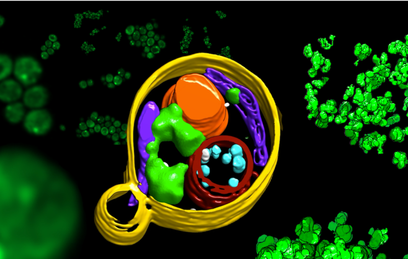
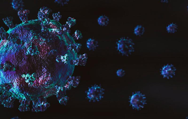
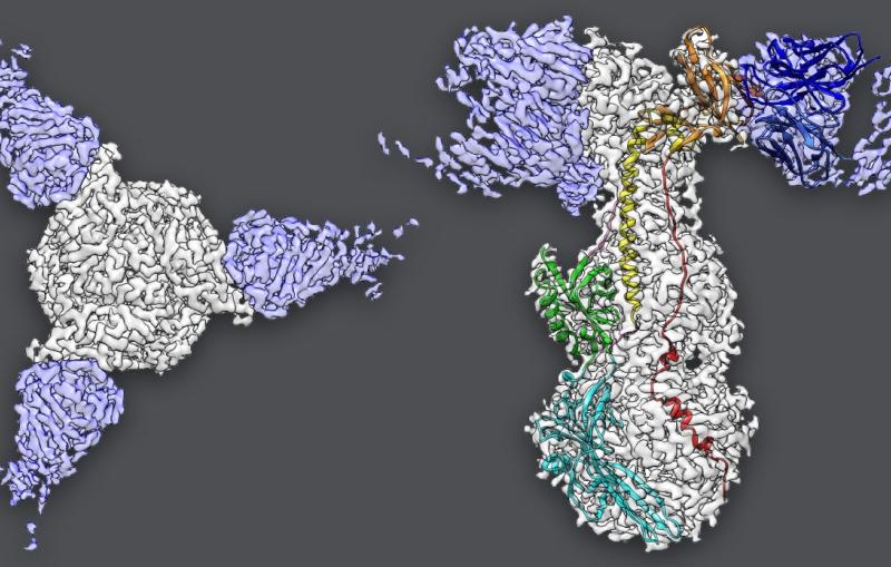
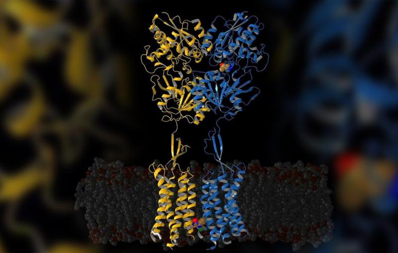
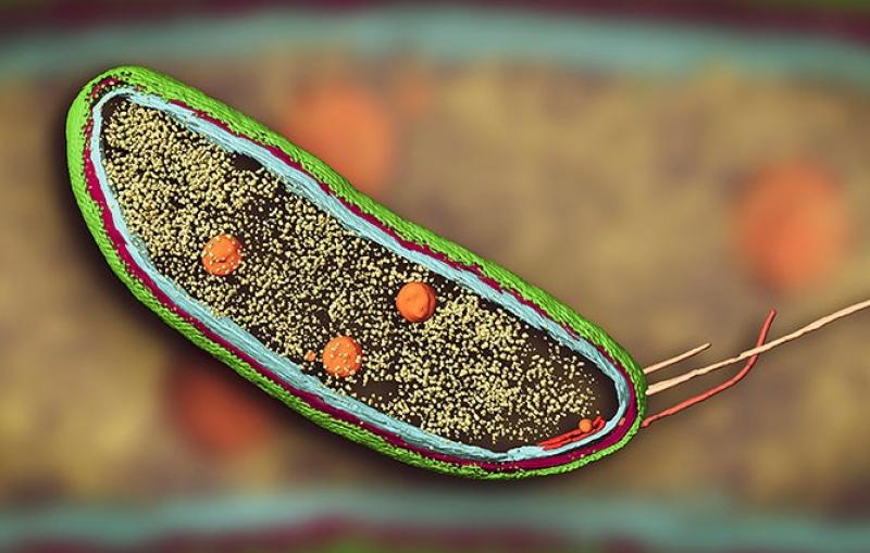
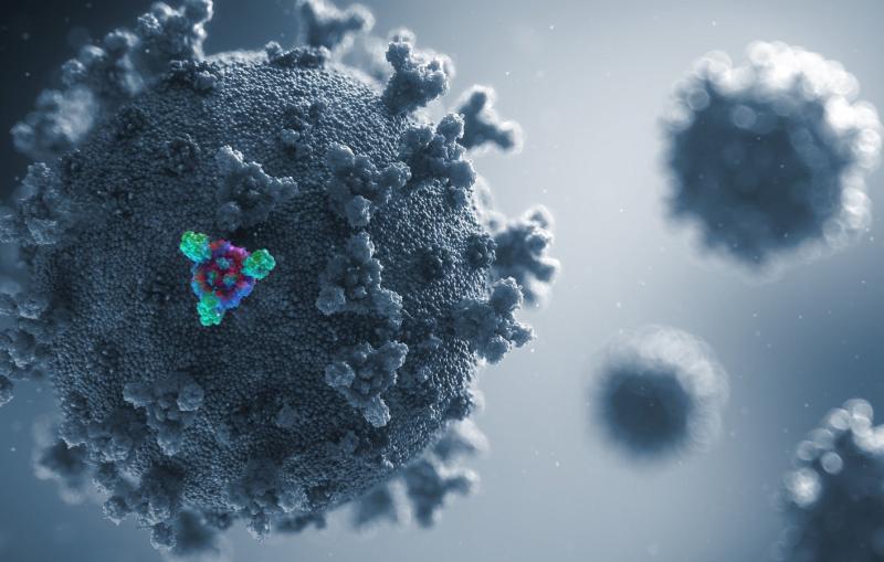
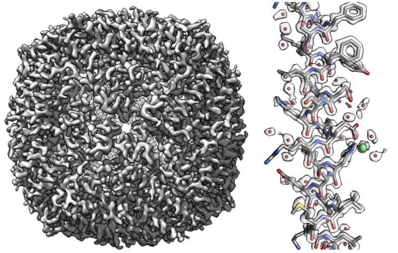
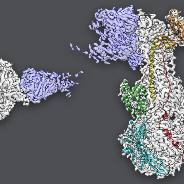
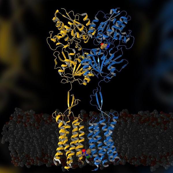
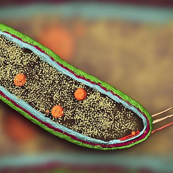
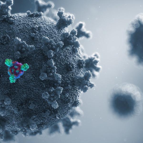
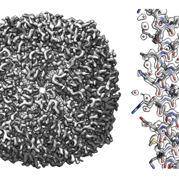
Cryo-EM allows scientists to make detailed 3D images of DNA, RNA, proteins, viruses, cells and the tiny molecular machines within the cell, revealing how they change shape and interact in complex ways while carrying out life’s functions.

Related links:   
Joint institutes and centers  
Cryo-EM fact sheet (pdf) 
Stanford-SLAC Cryo-Electron Microscopy website

Browse tagged content

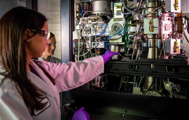
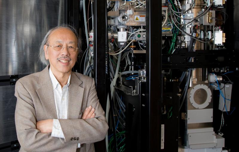
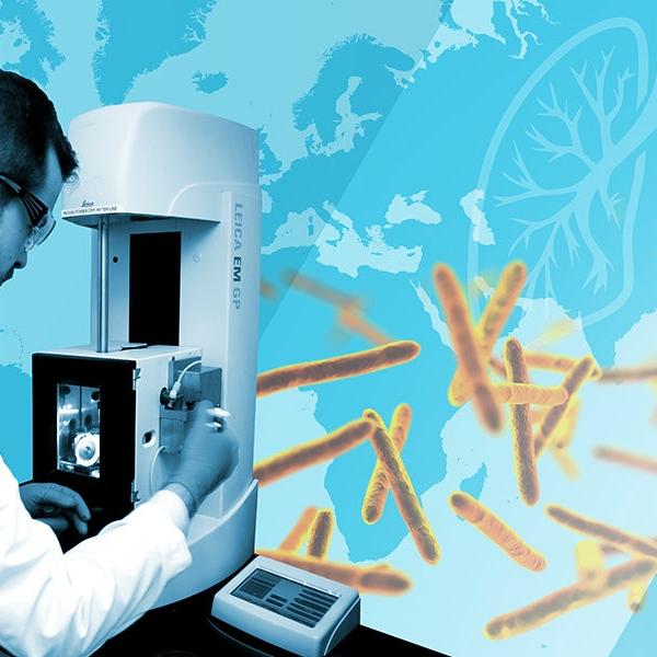
Research associate Megan Mayer and graduate student Patrick Mitchell load a sample into a cryogenic electron microscope at SLAC.
News Brief

The center complements other NIH centers at SLAC and elsewhere that broaden access to this cutting-edge technology for biomedical research.

Cryo-EM images of yeast cells with contents highlighted
News Brief

Images reveal how some antibodies may block SARS-CoV-2 infection.

A rendering of the SARS-CoV-2 virus.
Feature

The annual conference for scientists who conduct research at SLAC’s light sources engaged more than 1,700 researchers in talks, workshops and discussions.

2020 SSRL/LCLS Users' Meeting
Feature

No human cell can function without these tiny machines, which cause disease when they go haywire and offer potential targets for therapeutic drugs.

Illustration of molecular Ferris wheel moving protons
News Brief

Stanford and SLAC scientists studying the varicella zoster virus found that an antibody that blocks infection doesn’t work exactly as they’d thought.

Images extracted from cryo-EM data
Feature

A pioneer in developing methods for cryogenic electron microscopy, he directs two joint facilities for cryo-EM research and development on the SLAC campus.

Photo of Professor Wah Chui with a cryo-electron micrcoscope
News Brief

For the first time, scientists have revealed the steps needed to turn on a receptor that helps regulate neuron firing. The findings might help...

yellow and blue protein structures.
Feature

Researchers expect the new method to answer fundamental questions in biology and materials science. First up: Images showing molecules that help guide cell division...

cryo-EM image of Caulobacter bacterium
Feature

The lab is responding to the coronavirus crisis by imaging disease-related biomolecules, developing standards for reliable coronavirus testing and enabling other essential research.

SARS-CoV-2
News Release

The giant cavity, in a protein that transports nutrients across the cell membrane, is unlike anything researchers have seen before.

A scientist working overlaid on a world map and images of tuberculosis bacteria.
News Brief

Cryogenic electron microscopy can in principle make out individual atoms in a molecule, but distinguishing the crisp from the blurry parts of an image...

An overall image of the apoferritin molecule (left) and a small section (right)
News Brief

A new understanding of the nucleation process could shed light on how the shells help microbes interact with their environments, and help people design...

Illustration of tiles forming a microbial shell