Scientists marry two powerful techniques to pinpoint locations of individual molecules in their cellular neighborhoods
Researchers expect the new method to answer fundamental questions in biology and materials science. First up: Images showing molecules that help guide cell division in bacteria.
By Glennda Chui
Scientists have married two of today’s most powerful microscopy techniques to make images that pinpoint, for the first time, the identities and precise locations of individual proteins within the detailed context of bacterial cells. This information is crucial for learning how protein molecules work together to organize cell division and carry out other important tasks, such as enabling microbes to sniff out food and danger.
The new method has already unearthed new information about bacterial proteins and their nearby cellular neighborhoods. Researchers say it also has potential to answer fundamental questions about the molecular machinery of viruses, parasites, and processes like photosynthesis.
“This is a big leap for biology, and I think there are many, many systems that will benefit from this kind of imaging,” said Stanford Professor Lucy Shapiro, whose research group participated in the study.
The new hybrid method, called correlated imaging by annotation with single molecules, or CIASM (pronounced “chasm”), was developed by Peter Dahlberg, a postdoctoral researcher in the lab of Professor W. E. Moerner at Stanford University.
It’s a variation on a technique called low temperature single-molecule microscopy, invented by Moerner three decades ago, which attaches glowing labels to molecules so they can be individually identified. This method underlies super-resolution fluorescence microscopy, the topic of Moerner’s 2014 Nobel Prize in Chemistry.
What Dahlberg did was find a way to make this type of fluorescence imaging work at sub-freezing temperatures so the same samples could also be examined with cryogenic electron tomography (CET). CET uses streams of electrons to make 3D images of flash-frozen cells and their components at near-atomic resolution. Combining CET with the fluorescent imaging allows scientists to see the tagged molecules in the context of the surrounding cell, a crucial perspective for understanding their role in the cellular machinery.
“We can label specific molecules of interest so that the light we see comes only from those molecules, and then we find where they are within about 10 nanometers, or billionths of a meter. This gives us a much more accurate picture of what’s going on,” Dahlberg said. “We have taken the ultra-precise snapshots provided by CET and added a little bit of color.”
He added, “It is exciting to develop new imaging methods. When you are done, you get to take a step back and look at all the new questions you can attack.”
With CIASM, the research team was able to pinpoint the locations of three types of proteins in high-resolution CET images of bacteria taken at the Department of Energy’s SLAC National Accelerator Laboratory. The results were reported today in the Proceedings of the National Academy of Sciences.
“Every method has its advantages and disadvantages,“ Moerner said, “and this is a nice situation where we can combine two methods to learn more.”
Finding order in a cellular soup
Even in relatively simple bacterial cells, location is everything, said Saumya Saurabh, a postdoctoral researcher in Shapiro’s lab who played a leading role in the research.
“People tend to think of bacteria as sacks of proteins with no organization,” he said. “But it turns out that’s not true, and in fact many of the molecules in bacteria are precisely located in both space and time. If they’re not in the right position, the cell dies. What Pete’s work is finally allowing us to do is look inside with molecular resolution and find out when and where these molecules are located with respect to each other.”
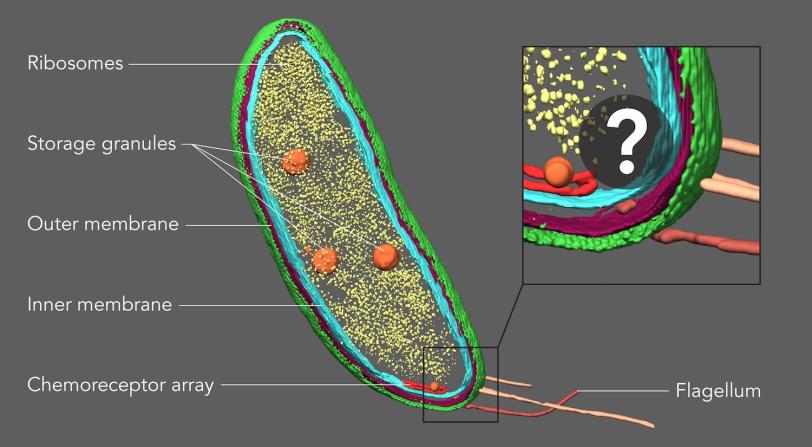
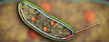
Caulobacter crescentus, for instance, a well-studied species of freshwater bacteria, is known for dividing into two very different types of daughter cells: One swims freely, while the other forms a stalk and attaches to a surface. How each daughter cell gets what it needs to follow its unique path has been a longstanding mystery.
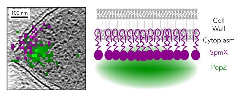
Scientists had previously identified small areas at either end of the dividing cell that might contain proteins that play key roles in this lopsided cell division. One of the proteins, PopZ, is found at both ends of the dividing cell, while the other, SpmX (“Spam-X”) is found only in the half that will develop a stalk.
For this study, Saurabh and graduate student Jiarui Wang labeled proteins in Caulobacter with fluorescent tags. Then Dahlberg froze these samples, performed single-molecule fluorescence imaging on them with the help of graduate student Annina Sartor, and took them to the Stanford-SLAC Cryo-EM facilities for CET imaging co-directed by Wah Chiu, a professor at Stanford and SLAC.
Mapping a protein hangout
The combined images not only confirmed that both proteins were in the areas scientists had suspected, but also revealed exactly how they were arranged: SpmX was embedded in the cell’s inner membrane and protruded into the cell’s interior, where it came into direct contact with PopZ.
“The exact orientation of this protein complex has been debated over the last 12 years,” Saurabh said. “We were able to observe the protein partners with exquisite resolution. Now we have a very precise picture of how these proteins talk to each other in the cell.”
The team tested the accuracy of CIASM by using it to confirm the location of a protein called McpA that was known to be part of a chemoreceptor array in the bacteria. “Exquisitely sensitive proteins in this array serve as Caulobacter’s nose,” Saurabh said, “sensing the chemistry of the surrounding environment so they can move away from unpleasant things and move toward the glucose they eat.”
The array appears as parallel black lines in CET images, and fluorescent tagging of the same images pinpointed the locations of individual McpA proteins within about 10 nanometers.
A detailed look at quantum dots
In a separate, parallel study, published in Angewandte Chemie on April 24, the researchers used a similar technique to look at single quantum dots, with some surprising results.
Quantum dots are nanoscale crystals of semiconductor material that naturally fluoresce in colors determined by their size, shape and composition. These dots are used in research to label and track proteins and other biological materials, and have potential applications in future electronics, lighting, quantum computing, medical imaging and other areas.
In this study, the goal was to see how the finer structural details of individual dots were related to specific details of their optical properties, said Davis Perez, a PhD student in Moerner’s lab.
“We were able to see some surprising behaviors of the individual quantum dots – for instance, in their response to excitation with laser light,” he said. “But the most exciting aspect to me is that the method we developed to study quantum dots can also be used to study biological systems such as photosynthetic proteins, where energy is transferred between groups of proteins, and see how the photosynthetic machinery operates.”
Moerner said his lab is working with Chiu to pursue these challenges.
“It is the early days of combining the two methods, and we are excited to explore more collaborations linking light and electrons,” Chiu said. “This hybrid imaging approach has the potential to uncover structures of molecular components involved in key biological processes in cells spanning all domains of life.”
Fluorescence microscopy for this study was carried out in the Moerner lab at Stanford.
The research was supported, in part, by the National Institute of General Medical Sciences and the Department of Energy Office of Science. The Stanford-SLAC Cryo-EM facilities were established through funding by Stanford University and SLAC National Accelerator Laboratory.
Citations
Peter D. Dahlberg et al., Proceedings of the National Academy of Sciences, 8 June 2020 (10.1073/pnas.2001849117)
Peter D. Dahlberg et al., Angewandte Chemie, 24 April 2020 (10.1002/anie.202002856)
Contact
For questions or comments, contact the SLAC Office of Communications at communications@slac.stanford.edu.
SLAC is a vibrant multiprogram laboratory that explores how the universe works at the biggest, smallest and fastest scales and invents powerful tools used by scientists around the globe. With research spanning particle physics, astrophysics and cosmology, materials, chemistry, bio- and energy sciences and scientific computing, we help solve real-world problems and advance the interests of the nation.
SLAC is operated by Stanford University for the U.S. Department of Energy’s Office of Science. The Office of Science is the single largest supporter of basic research in the physical sciences in the United States and is working to address some of the most pressing challenges of our time.
Related stories


Jennifer Cochran is the new Stanford vice president for SLAC and for strategic initiatives


