Compact Light Source Improves CT Scans
New Technology May Advance Preclinical Studies of Cancer and Other Diseases
A new study shows that the recently developed Compact Light Source (CLS) – a commercial X-ray source with roots in research and development efforts at the Department of Energy’s SLAC National Accelerator Laboratory – enables computer tomography scans that reveal more detail than routine scans performed at hospitals today. The new technology could soon be used in preclinical studies and help researchers better understand cancer and other diseases.
With its ability to image cross sections of the human body, X-ray computer tomography (CT) has become an important diagnostic tool in medicine. Conventional CT scans are very detailed when it comes to bones and other dense body parts that strongly absorb X-rays. However, the technique struggles with the visualization and distinction of “soft tissues” such as organs, which are more transparent to X-rays.
“Our work demonstrates that we can achieve better results with the Compact Light Source,” says Professor for Biomedical Physics Franz Pfeiffer of the Technical University of Munich in Germany, who led the new study published April 20 in the Proceedings of the National Academy of Sciences. “The CLS allows us to do multimodal tomography scans – a more advanced approach to X-ray imaging.”
More than One Kind of Contrast
The amount of detail in a CT scan depends on the difference in brightness, or contrast, which makes one type of tissue distinguishable from another. The absorption of X-rays – the basis for standard CT – is only one way to create contrast.
Alternatively, contrast can be generated from differences in how tissues change the direction of incoming X-rays, either through bending or scattering X-ray light. These techniques are known as phase-contrast and dark-field CT, respectively.
“Organs and other soft tissues don’t have a large absorption contrast, but they become visible in phase-contrast tomography,” says the study’s lead author, Elena Eggl, a researcher at the Technical University of Munich. “The dark-field method, on the other hand, is particularly sensitive to structures like vertebrae and the lung’s alveoli.”
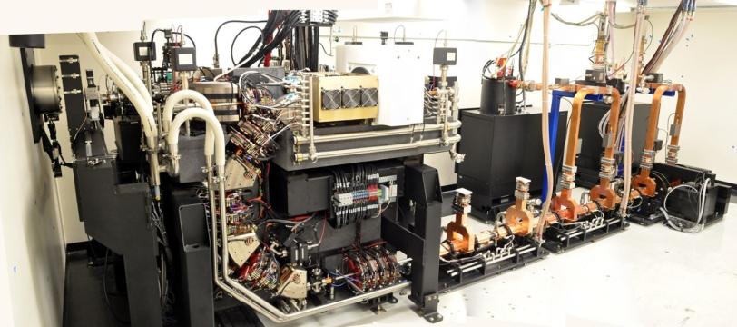
Shrinking the Synchrotron
However, these methods require X-ray light with a well-defined wavelength aligned in a particular way – properties that conventional CT scanners in hospitals do not deliver sufficiently.
For high-quality phase-contrast and dark-field imaging, researchers can use synchrotrons – dedicated facilities where electrons run laps in football-stadium-sized storage rings to produce the desired radiation – but these are large and expensive machines that cannot simply be implemented at every research institute and clinic.
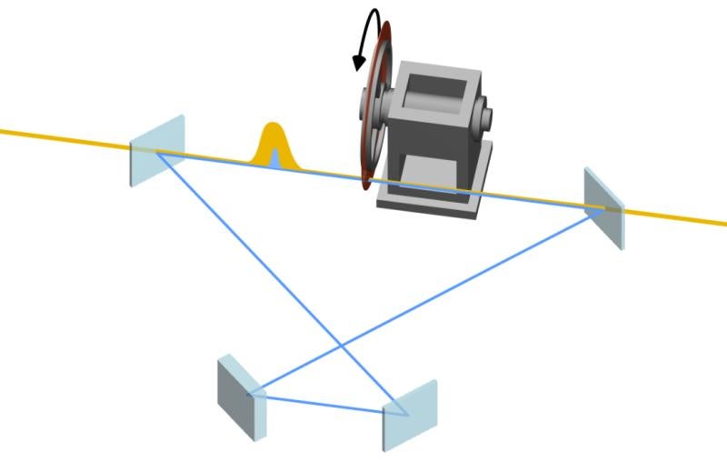
Conversely, the CLS is a miniature version of a synchrotron that produces suitable X-rays by colliding laser light with electrons circulating in a desk-sized storage ring. Due to its small footprint and lower cost, it could be operated in almost any location.
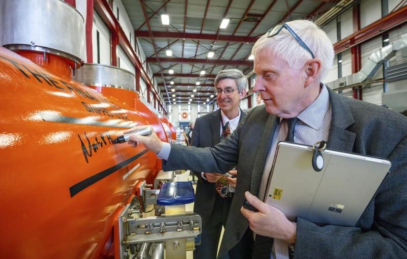
“The Large Hadron Collider at CERN is the world’s largest colliding beam storage ring, and the CLS is the smallest,” says SLAC scientist Ronald Ruth, one of the study’s co-authors. Ruth is also chairman of the board of directors and co-founder of Palo Alto-based Lyncean Technologies Inc., which developed the X-ray source based on earlier fundamental research at SLAC. “It turns out that the properties of the CLS are perfect for applications like tomography.”
More Modes, Finer Detail
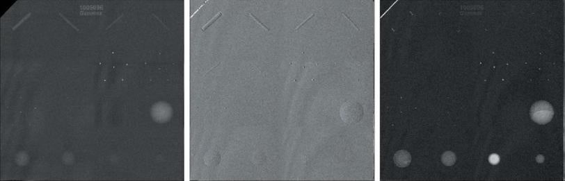
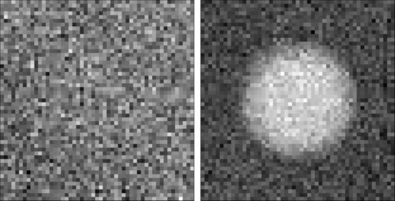
In the recent study, the researchers reported the first “multimodal” CT scan with the CLS: They recorded all three imaging modes – absorption, phase contrast and dark field – at the same time. Using a total of 361 two-dimensional X-ray images of an infant mouse taken from different directions, the scientists generated cross-section images of the animal.
“The absorption images only show bones and air-filled organs,” Eggl says. “However, the phase-contrast and dark-field images reveal much more detail, showing different organs such as the heart and liver. We can even distinguish different types of fat tissue, which is not possible with absorption-based CT scans.”
Using a standard sample of chemically well-defined liquids, the scientists also demonstrated that they could not only visualize but also quantify differences in their properties – information that can be applied to various body tissues and that is only obtained when combining all three tomography modes.
Implications for Cancer, Materials
The success of this research, which was done on a CLS prototype, has led to the commissioning of the first commercial device.
The researchers’ next goal is to use the CLS for phase-contrast and dark-field CT in preclinical studies – an approach that could help visualize cancer. “We work closely together with two clinics to study tumors,” Eggl says. “One of our plans is to image breast tissue samples and also entire breasts after mastectomy to better understand the clinical picture of breast cancer.”
Besides medical applications, multimodal tomography could also open up new possibilities in materials science, for instance, in studies of extremely durable and light-weight carbon fibers and other fibrous materials, where the X-ray absorption contrast provides little information.
Citation: E. Eggl, et al., Proceedings of the National Academy of Sciences of the United States of America, 20 April 2015 (10.1073/pnas.1500938112).
Further Information: Press release by the Technical University of Munich, Germany (30 April 2015).
For questions or comments, contact the SLAC Office of Communications at communications@slac.stanford.edu.
SLAC is a multi-program laboratory exploring frontier questions in photon science, astrophysics, particle physics and accelerator research. Located in Menlo Park, Calif., SLAC is operated by Stanford University for the U.S. Department of Energy's Office of Science.
SLAC National Accelerator Laboratory is supported by the Office of Science of the U.S. Department of Energy. The Office of Science is the single largest supporter of basic research in the physical sciences in the United States, and is working to address some of the most pressing challenges of our time. For more information, please visit science.energy.gov.
Related stories