Journal Highlights X-ray Laser Impacts on Biological Science
Even in their infancy, X-ray lasers such as SLAC's Linac Coherent Light Source are notching a list of important discoveries, and a special issue of a scientific journal highlights their unique contributions to biological sciences.
By Glenn Roberts Jr.
Even in their infancy, X-ray lasers such as SLAC's Linac Coherent Light Source (LCLS) are notching a list of important discoveries. A special issue of a scientific journal highlights their unique contributions to biological sciences and details technical milestones that are likely to secure their growing role in these fields.
LCLS, which started up in 2009 and is one of just three X-ray free-electron lasers in operation, has explored photosynthesis in action, membrane proteins that are key to drug discovery, viruses and other biologically important samples in ways not possible with more conventional tools.
"I don't think any of us would have predicted how quickly these lasers would be used to reveal new biology, and how inventive and diverse the science at these sources would be," said Henry Chapman, an X-ray laser pioneer at Germany’s DESY lab who co-edited the special issue of Philosophical Transactions of the Royal Society B, "Biology with free-electron X-ray lasers.” It appeared online June 9.
"When we look back at the initial list of experiments made before the LCLS was built, it seems we weren't dreaming big enough," Chapman said, "but once the machines turned on it stimulated the community to think of new possibilities."
His co-editor, John Spence of Arizona State University, is director of science for a new National Science Foundation Science and Technology Center, BioXFEL, which is focused on the application of X-ray lasers to biology. "We've seen many applications for X-ray laser research in biology, from drug design and the study of enzymes to photosynthesis and virology,” he said.
The issue features 26 scientific papers submitted by speakers who participated in BioXFEL’s first international conference, which was hosted by The Royal Society in October 2013.
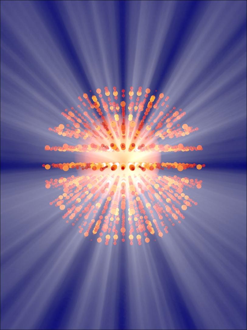
LCLS X-ray light is millions of times brighter than that of synchrotrons, its most advanced predecessors. X-ray lasers can study the atomic details of tiny biological samples in lifelike settings, and their light can capture changes measured in just quadrillionths of a second.
The extreme light produced by X-ray lasers "is the difference between a casual walk and traveling at the speed of light," said Richard Neutze of the University of Gothenburg in Sweden, whose paper highlights the opportunity to use X-ray lasers to study structural changes in proteins.
Among the other papers featured in the issue:
- Aina Cohen of SLAC's Stanford Synchrotron Radiation Lightsource (SSRL) and collaborators report on the use of an electron beam to produce detailed images of delicate crystallized biological samples used in LCLS experiments. The electron microscopy technique they describe can be used to view the quality of nanoscale crystals before and after they are jetted into a thin stream toward the X-ray laser pulses.
- A team led by Stanford and SLAC scientists report on a technique tested at SSRL that analyzes X-ray scattering patterns from a collection of tiny, randomly oriented particles to obtain atomic details about individual particles. Sebastian Doniach, a SLAC professor of photon science who participated in the study, said he hopes the technique, called correlated X-ray scattering, will become useful for X-ray laser studies of DNA, individual protein molecules and other biological samples.
- N. Duane Loh of the Stanford PULSE Institute, a joint institute of SLAC and Stanford, describes a theoretical technique, based on a data-crunching algorithm, to construct 3-D images of individual particles.
- Jan Kern of Lawrence Berkeley National Laboratory and SLAC highlights a suite of tools developed for use in X-ray laser studies of Photosystem II, an important protein complex in photosynthesis. These tools include a new type of liquid jet that sends crystallized samples of Photosystem II into the path of LCLS X-rays, devices that measure the chemistry of the samples, and software used to refine data from experiments.
Citation: Discussion Meeting Issue, "Biology with free-electron X-ray lasers," organized and edited by John C.H. Spence and Henry N. Chapman, Philosophical Transactions of The Royal Society B July 17, 2014; 369 (1647), DOI: 10.1098/rstb.2013.0500 1471-2970.
Contact
For questions or comments, contact the SLAC Office of Communications at communications@slac.stanford.edu.
SLAC is a multi-program laboratory exploring frontier questions in photon science, astrophysics, particle physics and accelerator research. Located in Menlo Park, California, SLAC is operated by Stanford University for the U.S. Department of Energy Office of Science.
SLAC’s LCLS is the world’s most powerful X-ray free-electron laser. A DOE Office of Science national user facility, its highly focused beam shines a billion times brighter than previous X-ray sources to shed light on fundamental processes of chemistry, materials and energy science, technology and life itself. For more information, visit lcls.slac.stanford.edu.
DOE’s Office of Science is the single largest supporter of basic research in the physical sciences in the United States, and is working to address some of the most pressing challenges of our time. For more information, please visit science.energy.gov.