SLAC topics

Cryo-EM RSS feed

Cryo-EM allows scientists to make detailed 3D images of DNA, RNA, proteins, viruses, cells and the tiny molecular machines within the cell, revealing how they change shape and interact in complex ways while carrying out life’s functions.

Related links:  
Joint institutes and centers 
Cryo-EM fact sheet 
Stanford-SLAC Cryo-Electron Microscopy website

Research associate Megan Mayer and graduate student Patrick Mitchell load a sample into a cryogenic electron microscope at SLAC.

Press Release

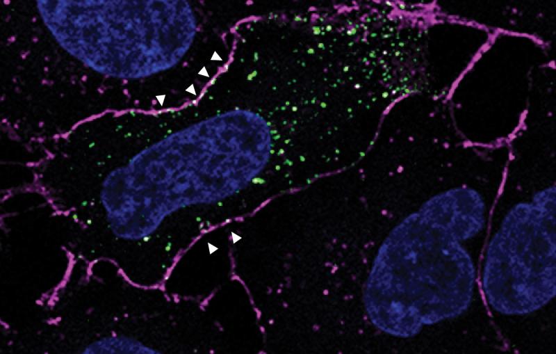
Researchers used cryo-EM (left) to discover how a chamber in human cells (right) directs protein folding. 

A pom-pom like object with curly tangles in purple and blue shades and yellow tangles at center, reminiscent of a zinnia blossom.
SLAC Science Explained

Taking pictures of tiny, flash-frozen things with electrons is revolutionizing biology and technology. SLAC and Stanford host one of the world’s leading facilities for...

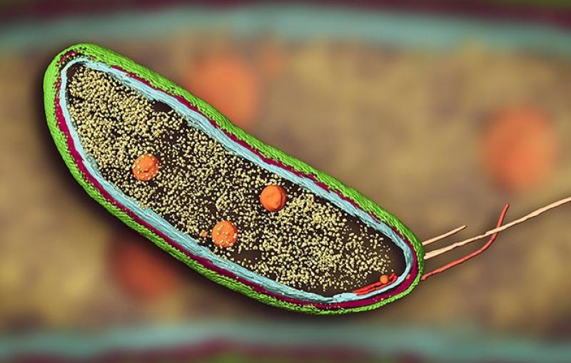
cryo-EM image of Caulobacter bacterium
News Feature

The leaders of SLAC's Technology Innovation Directorate discuss how their group supports the lab's most innovative projects.

TID senior managers
News Feature

SLAC’s Matt Garrett and Susan Simpkins talk about tech transfer that brings innovations from the national lab to the people, including advances for medical...

Tech Transfer
News Brief

Sandwiching wiggly proteins between two other layers allows scientists to get the most detailed images yet of a protein that’s key to the spread...

Red molecules are sandwiched between a blue inner shell and purple outer shell.
News Feature

In two new papers, researchers used X-ray crystallography and cryogenic electron microscopy to reveal new details of the structure and function of molecular assembly...

A model of the Lsd14 molecule
News Feature

Their work helps reveal the inner workings of cells and the behavior of matter under extreme pressures and temperatures.

News Feature

SLAC and Stanford scientists used it to zoom in on an iconic RNA catalyst and a piece of viral RNA that’s a potential target...

A high-res 3D ribbon diagram showing the structure of part of an RNA molecule
News Feature

From the invisible world of elementary particles to the mysteries of the cosmos, recipients of this prestigious award for early career scientists explore nature...

Panofsky fellows
News Feature

Scientists have documented a process that makes these next-gen batteries lose charge – and eventually some of their capacity for storing energy – even...

News Feature

Stanford EM-X brings hundreds of researchers around the world together to discuss the latest methods and discoveries from electron microscopes.

Black and white electron microscope images of pollen.
News Feature

G6PD deficiency affects about 400M people worldwide and can pose serious health risks. Uncovering the causes of the most severe cases could finally lead...