Clocking the movement of electrons inside an atom
Scientists get dramatically better resolution at X-ray free-electron lasers with a new technique.
Intense, ultrashort X-ray pulses from hard X-ray free-electron lasers (XFELs) can capture images of biological structures down to the atomic scale and shed light on the fastest processes in nature with a shutter speed of just one femtosecond, a millionth of a billionth of a second.
However, on these miniscule time scales, it is extremely difficult to synchronize the X-ray pulse that sparks a reaction in the sample with the follow-up pulse that observes the reaction. This problem, called timing jitter, is a major hurdle in performing these XFEL experiments with ever-better resolution.
Now, an international team that includes researchers from the Department of Energy’s SLAC National Accelerator Laboratory, the Max Planck Institute for the Structure and Dynamics of Matter, Deutsches Elektronen-Synchrotron Laboratory (DESY) and the Paul Scherrer Institute has found a way around this problem by measuring a fundamental decay process in neon gas at SLAC’s Linac Coherent Light Source (LCLS). The work was published in Nature Physics in January.
Shaking the jitters
Many biological systems – and some non-biological ones – suffer damage when they are excited by an X-ray pulse from an XFEL. One cause of damage is a process known as Auger decay: The X-ray pulse ejects some of the most tightly bound electrons, called photoelectrons, from atoms in the sample, and more weakly bound electrons fall in to take their place. This “relaxation” process releases energy and can induce the emission of yet another electron, known as an Auger electron.
Radiation from the intense X-rays and the continued emission of Auger electrons can rapidly damage the sample. In order to mitigate this damage, measurements ought to be captured before the decay sets in, so precise knowledge of the decay time scales is valuable. However, due to timing jitter, it is not generally possible to resolve such fast decay processes at XFELs.
“It’s like trying to photograph the end of a race when the camera shutter might activate at any moment in the final 10 seconds,” says lead author Dan Haynes, a doctoral student at Max Planck.
To circumvent the jitter problem, the research team came up with a highly precise way to chart Auger decay and demonstrated their method on samples of neon gas.
A streak of inspiration
The new technique builds upon established methods of streaking spectroscopy, where emitted electrons are accelerated or decelerated by the electric field of a “streaking” laser pulse. In this method, the XFEL pulse kickstarts the processes and the streaking pulse acts as a probe to observe them. Ordinarily, timing jitter restricts the resolution of this technique at XFELs.
After exposing both photoelectrons and Auger electrons to an external streaking laser pulse, the researchers determined their final kinetic energy in each of tens of thousands of individual measurements. Since Auger electrons are emitted later than photoelectrons, they also interact with the streaking laser pulse slightly later, and this consistent difference allowed the researchers to distinguish the two types of electrons from each other.
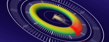
Despite the uncontrollable timing jitter between the two pulses, this consistent factor leads to a characteristic elliptical shape in the analyzed data when the measurements are plotted on a grid. The position of individual data points around the ellipse can be read like the hands of a clock to reveal the precise timing of ultrafast electron motions.
The researchers are hopeful that the technique will have a broader impact in the field of ultrafast science. In addition, Auger decay is a key factor in studies of exotic, highly excited states of matter, which can only be investigated at XFELs.
“This technique allowed us to measure the delay with sub-femtosecond precision, even though the timing jitter during the experiment was in the hundred-femtosecond range,” Haynes says, “It may facilitate a new class of experiments benefiting from the flexibility and extreme intensity of XFELs.”
LCLS is a DOE Office of Science user facility. This research was supported by the DOE Office of Science.
Citation: D.C. Haynes et al., Nature Physics, 18 January 2021 (10.1038/s41567-020-01111-0)
For questions or comments, contact the SLAC Office of Communications at communications@slac.stanford.edu.
Image caption: Precise synchronization of X-ray and external laser pulses is a major hurdle in analyzing data from XFEL experiments. Researchers took advantage of timing differences between two types of electrons, plotting tens of thousands measurements that formed a characteristic elliptical shape on a grid. Their results showed that the position of these individual data points can be read like the hands of a clock to reveal the timing of ultrafast electron motions. (Daniel Haynes/Jörg Harms)
SLAC is a vibrant multiprogram laboratory that explores how the universe works at the biggest, smallest and fastest scales and invents powerful tools used by scientists around the globe. With research spanning particle physics, astrophysics and cosmology, materials, chemistry, bio- and energy sciences and scientific computing, we help solve real-world problems and advance the interests of the nation.
SLAC is operated by Stanford University for the U.S. Department of Energy’s Office of Science. The Office of Science is the single largest supporter of basic research in the physical sciences in the United States and is working to address some of the most pressing challenges of our time.


