In a first, researchers use ultrafast ‘electron camera’ to learn about molecules in liquid samples
This new technology could enable future insights into chemical and biological processes that occur in solution, such as vision, catalysis and photosynthesis.
High-speed “electron cameras” can detect tiny molecular movements in a material by scattering a powerful beam of electrons off a sample. Until recently, researchers had only used this technique to study gases and solids. But some of the most important biological and chemical processes, in particular the conversion of light into energy, happen in molecules in a solution.
Now, researchers have applied this technique, ultrafast electron diffraction, to molecules in liquid samples. They developed a method to create 100-nanometer thick liquid jets–about 1,000 times thinner than the width of a human hair–that enable them to get clear diffraction patterns from electrons. In the future, this method could allow them to explore light-driven processes such as vision, catalysis, photosynthesis and DNA damage caused by UV rays.
The team, which included researchers from the Department of Energy’s SLAC National Accelerator Laboratory, Stanford University and the University of Nebraska-Lincoln (UNL), published their results in Structural Dynamics in March.
“This research is a huge breakthrough in the field of ultrafast electron diffraction,” says Xijie Wang, director of the MeV-UED instrument, who co-authored the paper. “Being able to study biological and chemical systems in their natural environment is a valuable tool that opens up a new window for the future.”
Stop-motion movies
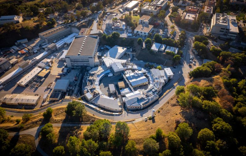
Liquid jets have long been used to deliver samples at X-ray lasers such as SLAC’s Linac Coherent Light Source (LCLS), providing valuable information about ultrafast processes as they occur in their natural environment. SLAC’s ultrafast “electron camera,” MeV-UED, uses high-energy electron beams to complement the range of structural information collected at LCLS.
Here, scientists begin by exciting a sample with laser light, kicking off the processes they hope to study. Next they blast the sample with a short pulse of electrons with high energy, measured in millions of electronvolts (MeV), to look inside, generating snapshots of its shifting atomic structure that can be strung together into a stop-motion movie of the light-induced structural changes in the sample.
Looking into the kaleidoscope
The tiny wavelengths of these high-energy electrons allow scientists to take high-resolution snapshots, offering insight into processes such as proton transfer and hydrogen-bond breaking that are difficult to study with other methods. But applying this technique to liquid samples has proven challenging.
“Since electrons don’t penetrate samples as easily as X-rays,” says Kathryn Ledbetter, a graduate student at the Stanford PULSE Institute who coauthored the paper, “applying this technique to liquids has been a longstanding challenge in the field.”
If the sample is too thick, the electrons can get stuck and scatter multiple times, producing a wild mix of patterns that’s difficult to glean information from, like looking through a kaleidoscope. In this new study, the team overcame those challenges through the use of MeV electrons and a gas-accelerated thin liquid sheet jet. As the electrons break through the jet, they scatter only once, producing a clean pattern that’s much easier to reconstruct. The team also designed a chamber that housed the liquid jet and monitored the interaction between the sample and the electron beam.
‘Another tool in the ultrafast toolbox’
This paper sets the stage for upcoming research that investigates questions such as what happens when hydrogen bonds break or when molecules absorb UV radiation. As a next step, SLAC researchers are upgrading the MeV-UED facility and developing a new generation of direct electron detectors that will greatly expand the scientific reach of this technique.
“We’d like this to be another tool in the toolbox of researchers trying to learn about liquids and light-driven reactions,” says Pedro Nunes, a postdoctoral researcher at UNL who led the research. “We want to show the community that what was once believed to be far-fetched is not only possible, but capable of running smoothly enough to watch structural changes unfold in real time.”
UED-MeV and LCLS are DOE Office of Science user facilities. The project was funded by the Office of Science.
Citation: J.P.F. Nunes et al., Structural Dynamics, 9 March 2020 (10.1063/1.5144518)
Contact
For questions or comments, contact the SLAC Office of Communications at communications@slac.stanford.edu.
SLAC is a vibrant multiprogram laboratory that explores how the universe works at the biggest, smallest and fastest scales and invents powerful tools used by scientists around the globe. With research spanning particle physics, astrophysics and cosmology, materials, chemistry, bio- and energy sciences and scientific computing, we help solve real-world problems and advance the interests of the nation.
SLAC is operated by Stanford University for the U.S. Department of Energy’s Office of Science. The Office of Science is the single largest supporter of basic research in the physical sciences in the United States and is working to address some of the most pressing challenges of our time.


