In brief: After blasting a molecule with light, researchers watch its structure vibrate and change in real time
The technique can be used to study molecular phenomena and the forming and breaking of chemical bonds.
A new study describes how a team of researchers watched a molecule vibrate after they excited it with ultraviolet light. The scientists were able to measure, with great clarity, how the structure of a molecule in an excited state changed over time. This research is driving a better understanding of how light directs molecular motions, which will enable the design of new chemical systems for a wide range of applications. The team, which includes researchers from Brown University, the Department of Energy’s SLAC National Accelerator Laboratory and the University of Edinburgh, UK, published their findings in Nature Chemistry last week.
Many chemical reactions happen extremely quickly, often in just millionths of a billionth of a second, making it difficult to track them as they’re occurring. Until recently, chemists relied on before-and-after shots of molecules involved in chemical reactions to learn more about how the reaction unfolded, or they used indirect methods from which they have to infer the molecular motion. Over the past decade, the development of ultrafast X-ray lasers has enabled scientists to measure chemical reactions at the atomic scale as they occur.
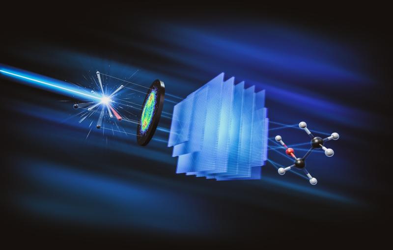
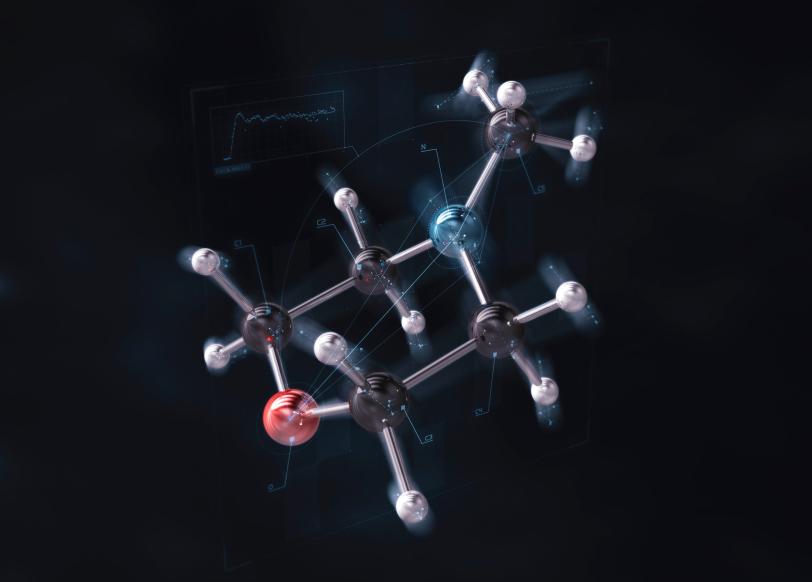
Using SLAC’s Linac Coherent Light Source X-ray laser (LCLS), the team scattered ultrafast pulses of X-rays off the molecule N-methyl morpholine after exciting it with UV light, creating snapshots of how it moved that were pieced together into a “molecular movie.” This allowed them to see how the electron cloud of the molecule rearranged and also measure the structure of the molecule as it vibrated. The research allowed the team to observe subtle effects in the organic molecule with a new level of clarity and revealed some surprising behavior in how the reaction evolved long after the initial excitation.
With the high sensitivity achieved in this paper, the researchers will be able to see changes in the arrangements of individual electrons in reacting molecules. The technique can be used to study molecular phenomena and the forming and breaking of chemical bonds as they happen, which will improve fundamental knowledge of chemistry.
The study was led by Brian Stankus and Haiwang Yong, researchers at Brown. Peter Weber, a professor of chemistry at Brown; Adam Kirrander, a professor of chemistry at the University of Edinburgh; and Mike Minitti, a senior staff scientist at SLAC, were the principal investigators. LCLS is a DOE Office of Science user facility. This research was supported by the Office of Science and the Army Research Office.
Citation: B. Stankus et al., Nature Chemistry, 8 July 2019 (10.1038/s41557-019-0291-0)
Contact
For questions or comments, contact the SLAC Office of Communications at communications@slac.stanford.edu.
SLAC is a vibrant multiprogram laboratory that explores how the universe works at the biggest, smallest and fastest scales and invents powerful tools used by scientists around the globe. With research spanning particle physics, astrophysics and cosmology, materials, chemistry, bio and energy sciences and scientific computing, we help solve real-world problems and advance the interests of the nation.
SLAC is operated by Stanford University for the U.S. Department of Energy’s Office of Science. The Office of Science is the single largest supporter of basic research in the physical sciences in the United States, and is working to address some of the most pressing challenges of our time. For more information, please visit science.energy.gov.