SLAC topics

X-ray light sources and electron imaging RSS feed

X-ray light sources and electron imaging are advanced techniques used to study the structure and properties of materials. X-ray light sources use high-energy photons to produce X-rays, while electron imaging uses high-energy electrons to produce detailed images of samples. 

Browse tagged content

Aerial view of SLAC
Feature

Scientists at SLAC and Utrecht University have identified how catalysts degrade when used to refine crude oil.

Image - Research at the Department of Energy’s SLAC National Accelerator Laboratory could lead to more efficient gasoline production. (@iStockphoto/Patryk Kosmider)
News Release

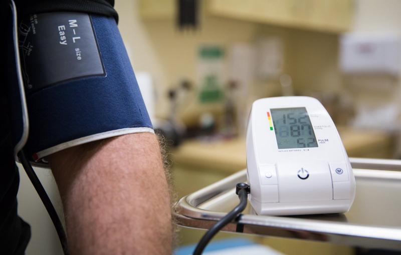
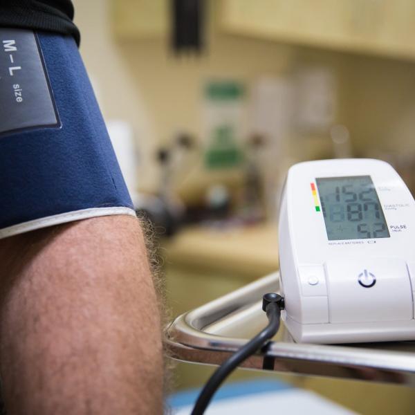
An experiment at SLAC's X-ray laser has revealed in atomic detail how a hypertension drug binds to a cellular receptor that plays a key...

Image - This photo shows a medical device used to monitor blood pressure. In a study at SLAC's Linac Coherent Light Source X-ray laser, researchers studied how a hypertension drug binds to a cellular receptor known as an angiotensin II type 1 receptor.
Feature

Stanford University and the Department of Energy’s SLAC National Accelerator Laboratory have established the first endowed professorship in honor of Arthur Bienenstock.

Feature

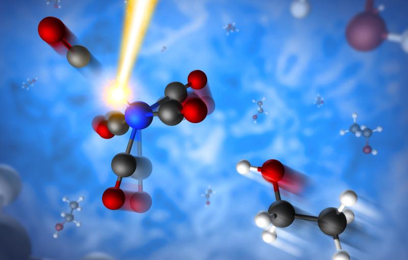
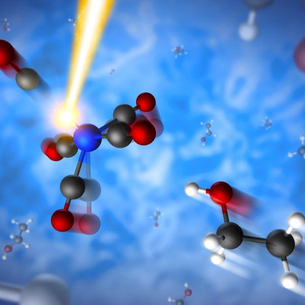
Researchers use X-ray laser at SLAC to track light-triggered chemical reactions in a molecule that serves as a simple model for the conversion of...

IMAGE - Artistic rendering of a molecule severed by laser light, with a separate molecule (bottom right) from a solvent rushing in to bond with the just-split molecule. (SLAC National Accelerator Laboratory)

Feature

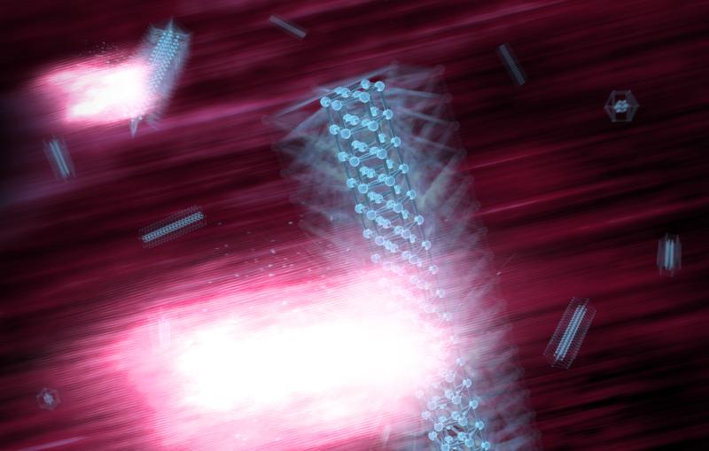
SLAC study of tiny nanocrystals provides new insight on the design and function of nanomaterials

Image - In this illustration, intense X-rays produced at SLAC's Linac Coherent Light Source strike nanowires to study an ultrafast "breathing" response in the crystals induced quadrillionths of a second earlier by pulses of optical laser light.
Feature

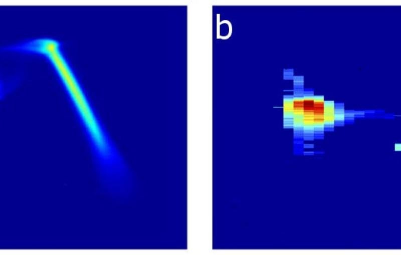
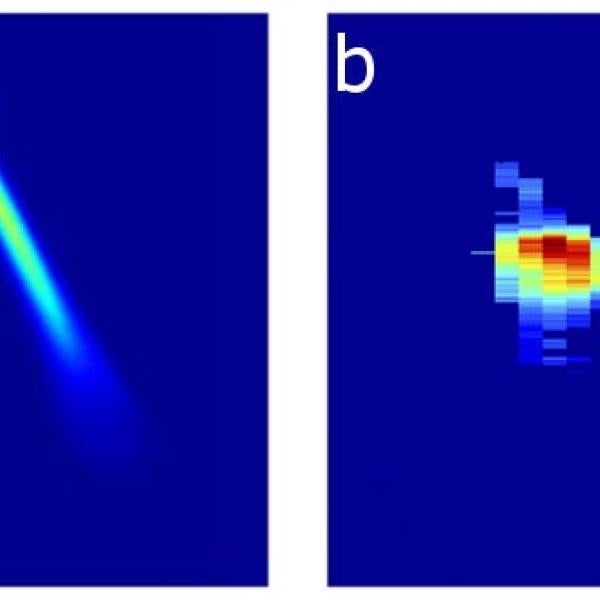
Developed at SLAC’s LCLS, it could also yield new information from hard-to-study samples in materials science, chemistry and other fields.

Image - These charts show (a) the energy profile of two electron bunches that are separated by about 6 picoseconds, which are later stimulated to emit (b) two X-ray pulses separated by femtoseconds.
Feature

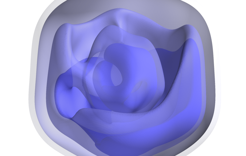
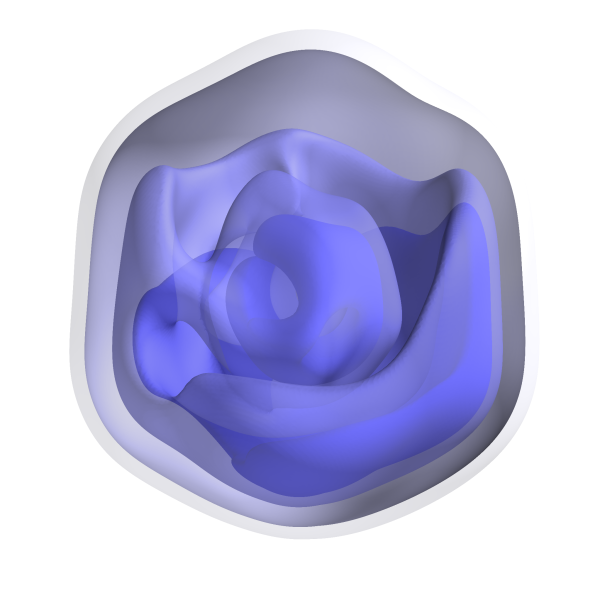
For the first time, researchers have produced a 3-D image revealing some of the inner structure of an intact, infectious virus.

Image - This rendering shows a 3-D reconstruction of a Mimivirus, based on an analysis of a collection of X-ray diffraction patterns obtained in an experiment at SLAC's Linac Coherent Light Source X-ray laser. (Uppsala University)
Feature

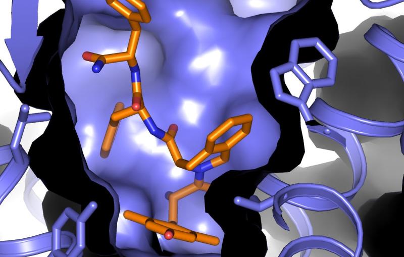
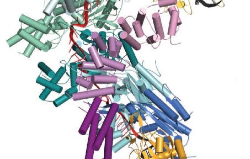
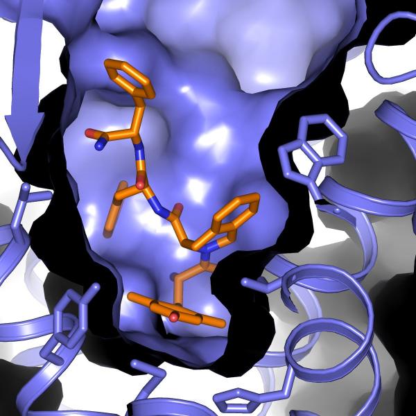
An X-ray laser experiment could lead to new drugs that lessen the side effects caused by powerful painkillers like morphine.

Image - This rendering shows a type of cellular membrane protein known as a delta opioid receptor (purple) with a compound derived from a naturally occurring peptide (orange, blue and red) bound inside its “pocket.” The peptide compound shows promise as a
News Release

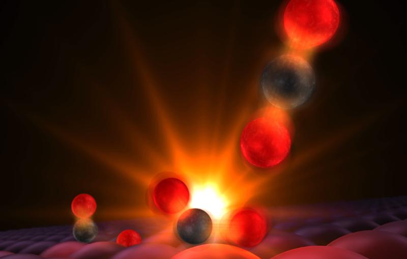
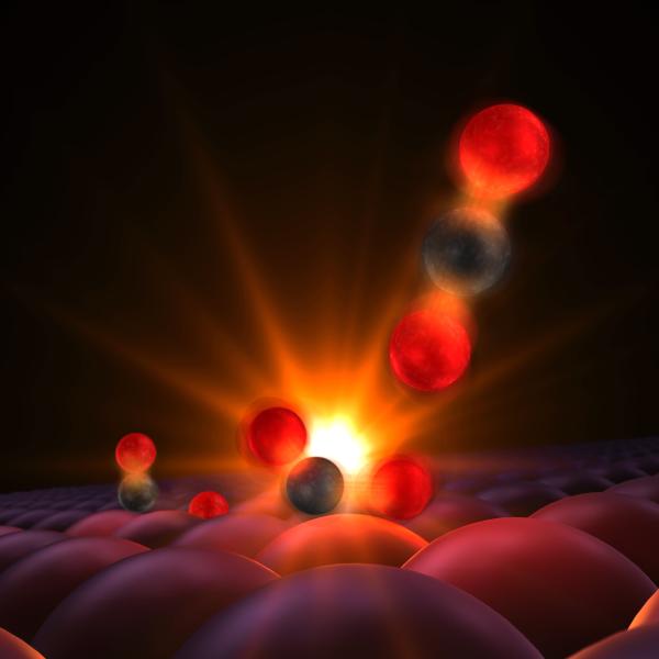
Scientists have used an X-ray laser at SLAC to get the first glimpse of the transition state where two atoms begin to form a...

Illustration of a transition state in a chemical reaction.
News Release

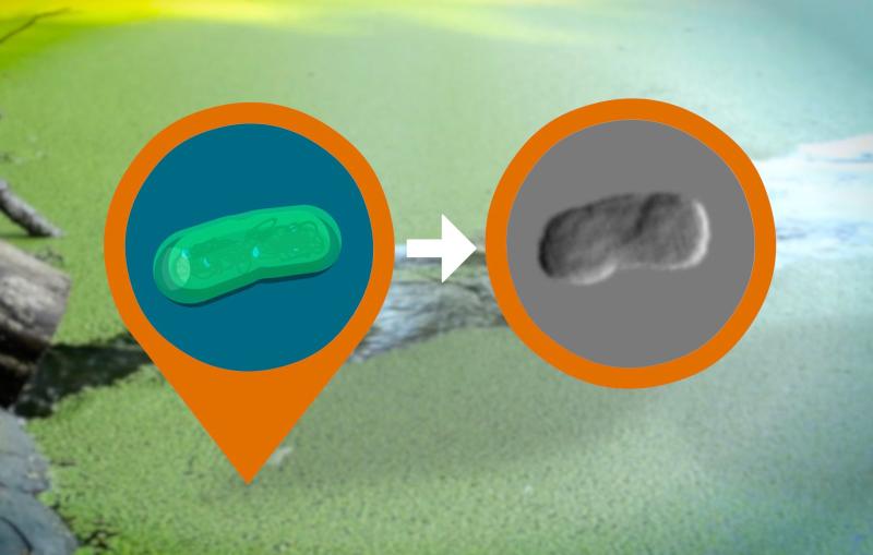
Technique Could Allow Study of Viral Infections, Cell Division and Photosynthesis in New Detail

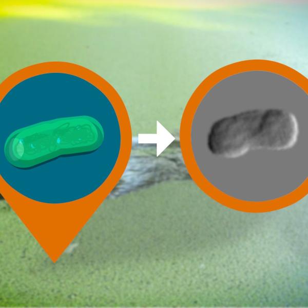
A pond containing a visible bloom of cyanobacteria, with an artistic rendering of an individual cell
Feature

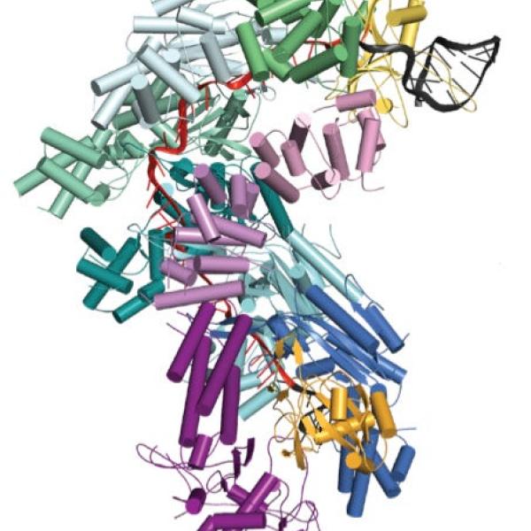
Research reveals that the bacterial immune system can better destroy viral attackers by saving genetic records of previously encountered viruses.

Feature

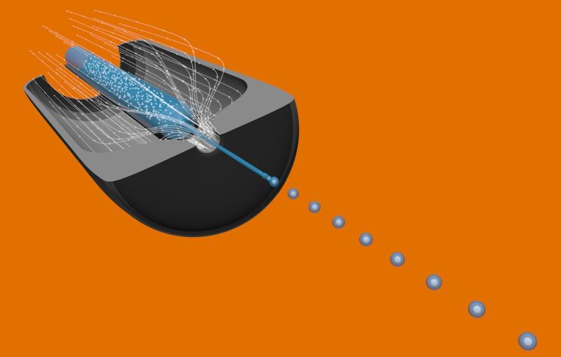
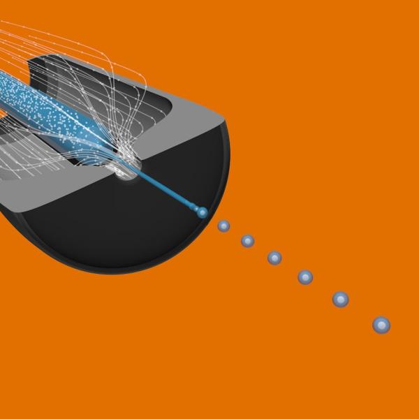
Scientists have assembled an exotic toolbox for experiments that tap into the brightest X-rays on the planet.

Image - This illustration shows a cutaway view of a type of sample system used at the Linac Coherent Light Source X-ray laser that jets samples in a superthin liquid or gel stream into its X-ray pulses. This system is known as a gas dynamic virtual nozzle