SLAC topics
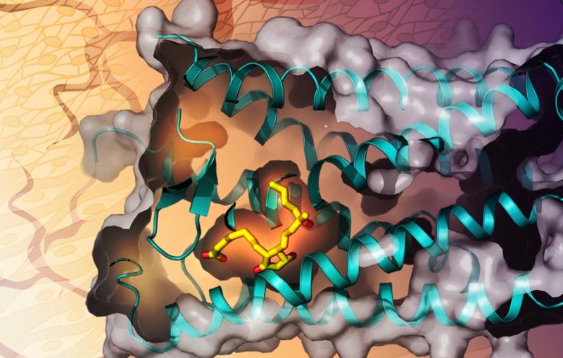
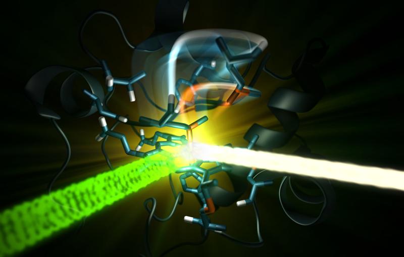
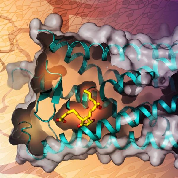
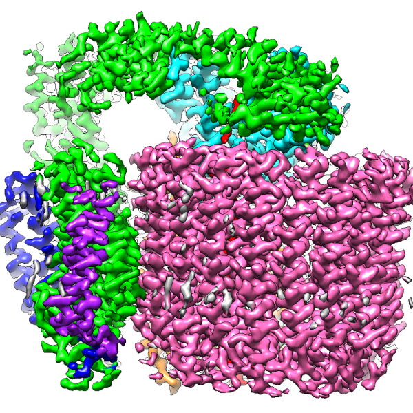
Structural molecular biology
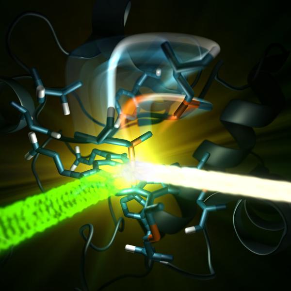
Structural molecular biology uses various scientific techniques to map the three-dimensional arrangement of atoms in biological molecules.
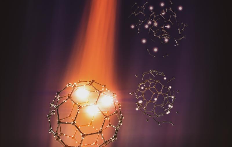
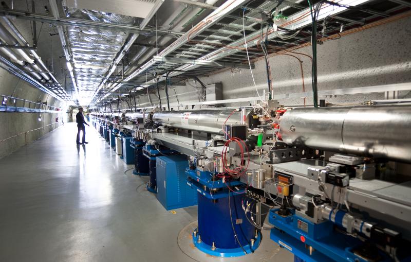
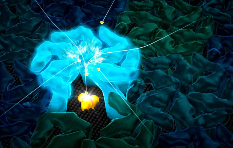
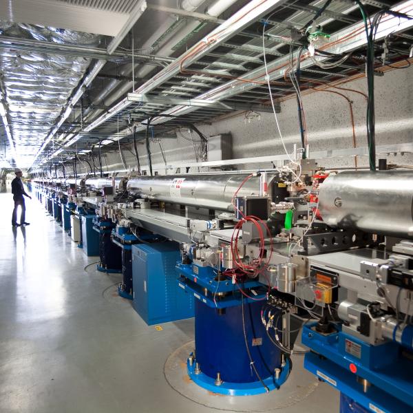
The past decade has seen the exciting birth of the first X-ray laser, the LCLS free electron laser (FEL) followed by other FELs around the world, leading to an explosion of new science, in the femtosecond and very recently in the attosecond regime. I will present our recent time-resolved experimental results using pump-probe technique with FELs to watch, in real time, the response of large molecules to intense X-rays as well as to examine the role of physical and chemical effects and how they lead to the timing of bonds breaking and bond forming.
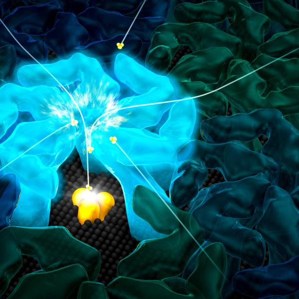
(Greg Stewart/SLAC National Accelerator Laboratory)