SLAC Will Open One of Three NIH National Service Centers for Cryo-Electron Microscopy
The National Institutes of Health center on the SLAC campus will make this revolutionary technology available to scientists nationwide and teach them how to use it to study 3D structures of biological machines and molecules.
By Glennda Chui
Menlo Park – The National Institutes of Health announced today that it will establish a national service and training center for cryogenic electron microscopy research at the Department of Energy’s SLAC National Accelerator Laboratory.
It’s one of three national service and training centers the NIH is setting up to make the Nobel prize-winning technology available to scientists nationwide and teach them how to use it.
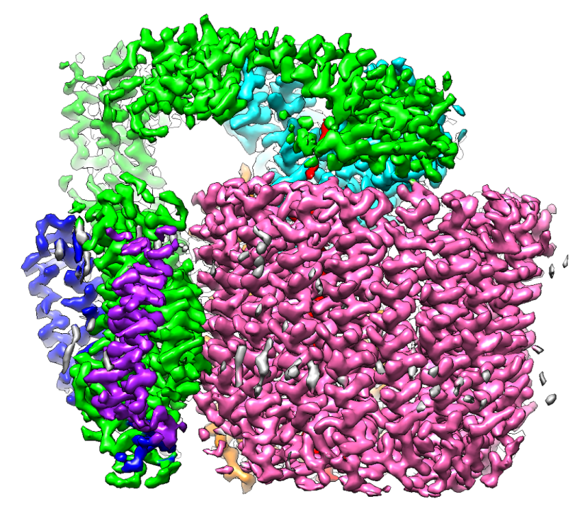
Known as cryo-EM for short, this powerful high-resolution imaging method has become a revolutionary tool for biology over the past few years due to rapid improvements in transmission electron microscopes, detectors and software. Last year the technique earned three of its key developers the 2017 Nobel Prize in chemistry. Cryo-EM allows scientists to make detailed 3D images of DNA, RNA, proteins, viruses, cells and the tiny molecular machines within the cell, revealing how they change shape and interact in complex ways while carrying out life’s functions.
However, the high cost of buying and operating the high-voltage electron microscopes and a lack of training opportunities have slowed the widespread adoption of the technology.
The new data collection centers will address this by providing funding for instruments and associated lab equipment and bringing in scientists from across the nation for research and training. In addition to SLAC, centers will be set up at the New York Structural Biology Center and at the Oregon Health & Science University in partnership with DOE’s Pacific Northwest National Laboratory, the NIH announced. The awards are anticipated to total $128 million over six years, pending the availability of funds.
“Cryo-electron microscopy is allowing us to resolve the three-dimensional structures of important biomolecules involved in disease that were inaccessible using previous technologies,” said National Institute of General Medical Sciences Director Jon R. Lorsch. “NIH wants to ensure as many scientists as possible have access to this crucial technology.”
The NIH center at SLAC will be known as the SLAC-Stanford Cryo-EM Center (S2C2). It marks the second major step in carrying out the Stanford-SLAC Cryo-EM Initiative, whose goal is to establish one of the world’s foremost hubs for cryo-EM research and training for scientists at the lab, the university and in the broader scientific community around the globe.
The first step took place earlier this year, when the Stanford-SLAC Cryo-EM Facility opened on the SLAC campus with four state-of-the-art microscopes.
The new NIH center, which will operate independently but in synergy with the recently established Stanford-SLAC facility, will install several more electron microscopes and associated specimen preparation devices in the lab’s soon-to-open Arrillaga Science Center.
“This new center complements our existing facilities and capabilities, enhancing an integrated suite of unique tools to advance materials, chemical and biological science discoveries critical to the DOE Office of Science mission,” said SLAC Director Chi-Chang Kao.
Wah Chiu, a professor at SLAC and Stanford and leader of the cryo-EM program, added, “This is an exciting moment for those in the U.S. scientific community who do not have access to cryo-EM instrumentation in their own institutions, and we are very pleased to share our decades of experience in this research with others.
“I believe that this NIH initiative will have a great impact on popularizing this powerful imaging tool,” he said, “which will likely lead to many discoveries of 3D structures of biological machines and molecules in both their normal and diseased states and hasten our national efforts to prevent and cure a variety of diseases, including cancer, diabetics, neurodegeneration, cardiovascular diseases and infection.”
Contact
For questions or comments, contact the SLAC Office of Communications at communications@slac.stanford.edu.
SLAC is a multi-program laboratory exploring frontier questions in photon science, astrophysics, particle physics and accelerator research. Located in Menlo Park, Calif., SLAC is operated by Stanford University for the U.S. Department of Energy's Office of Science.
Funding for S2C2 comes through Transformative High Resolution Cryo-Electron Microscopy program grant U24 GM-129541. The program is part of the NIH Common Fund, which encourages collaboration and supports a series of exceptionally high-impact, trans-NIH programs. Common Fund programs are managed by the Office of Strategic Coordination in the Division of Program Coordination, Planning, and Strategic Initiatives in the NIH Office of the Director in partnership with the NIH Institutes, Centers, and Offices.
SLAC National Accelerator Laboratory is supported by the Office of Science of the U.S. Department of Energy. The Office of Science is the single largest supporter of basic research in the physical sciences in the United States, and is working to address some of the most pressing challenges of our time.
Related stories


Jennifer Cochran is the new Stanford vice president for SLAC and for strategic initiatives


