

News Brief
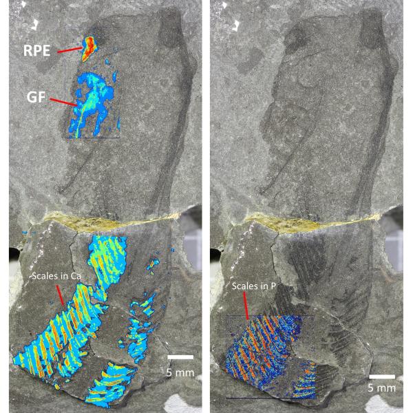
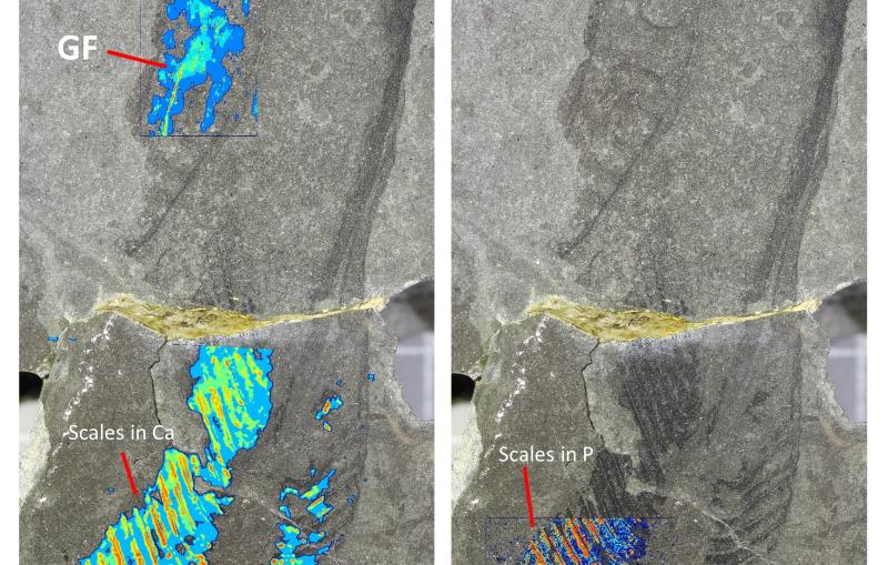
Via The University of Manchester
443-million-year-old fossils reveal early vertebrate eyes

News Brief
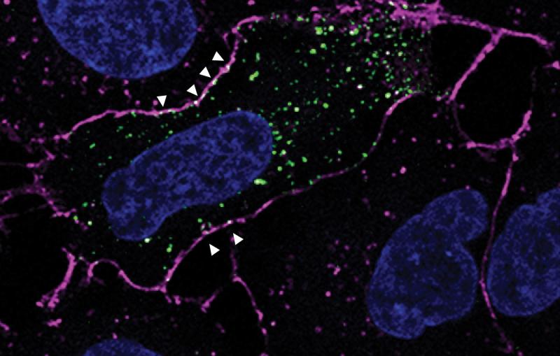
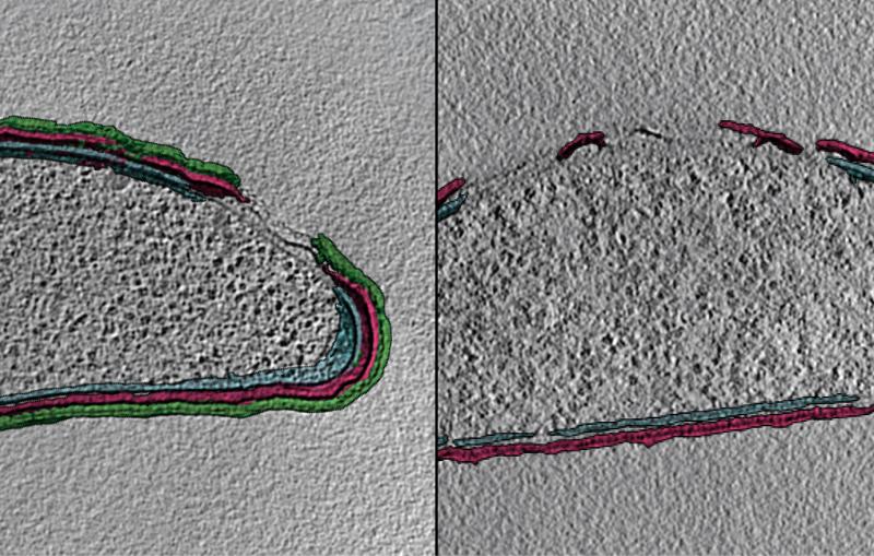
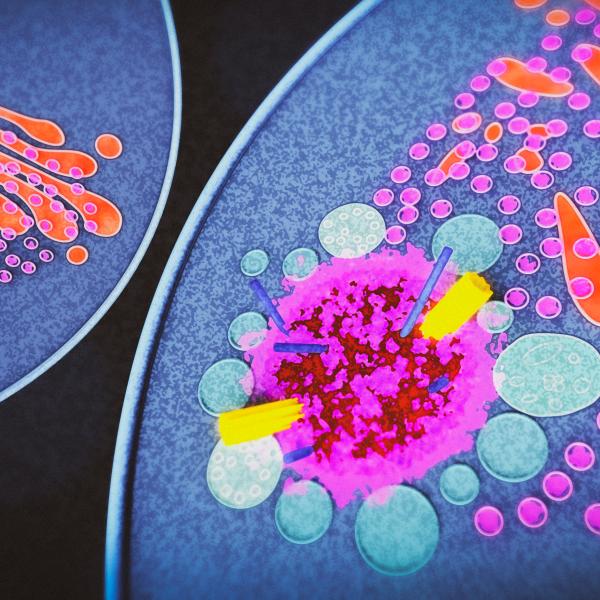
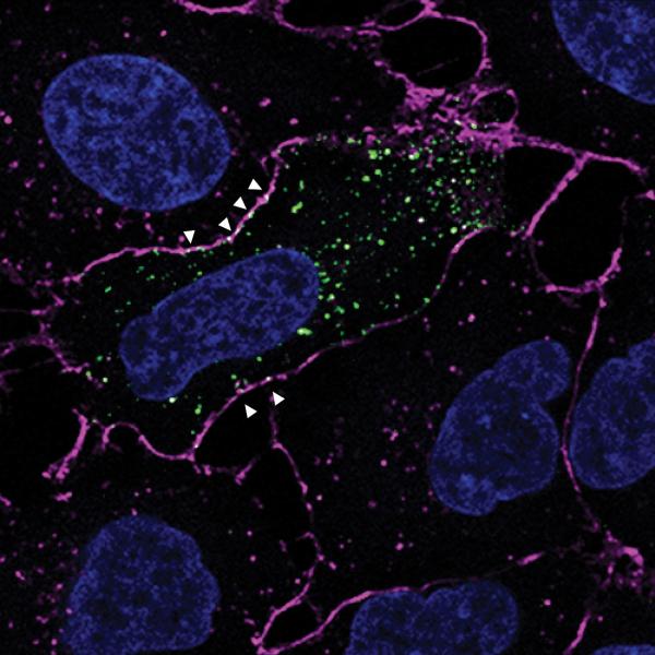
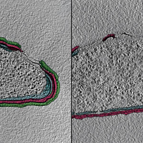
Via Innovative Genomics Institute
New method uncovers how viruses evade immune responses — and how we might fight back