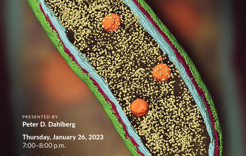
molecular machines they do all the work of life and professor watch who's been
studying them for many years first at Baylor University and then lately at SLAC facility who's developing
here on crow I am in my new detail he wants its concerns today how they are
put together how they work how they do all the work that life does so thank you
very much it's very nice of you to come out this evening to join me to listen to
the counter work I've been doing in the last 30 or 40 years and so I am a
professor at photon science here slack and also in the engineering in the
engineering school as well as the microbiology and immunology in the medical school at Stanford so first of
all I can't lie to make an introduction what do we mean by molecular machines and what is really quiet
em and so here is the work molecular machine being introduced by Professor
Bruce Abbott at University California in San Francisco Medical School and he draw
an analogy a molecular machine to a man-made machine as you can imagine a memory machine just an automobile is
made up of many parts each of this part has to work cooperative and is a dynamic
process is not this sitting still so a molecular machine in a living system is
also making a many parts but those power proteins and decay assets and all this
part has to put in the right place and has to move at the right time so
these are the challenge about how these molecular machines are being together
and how they function in the cell and I would say that there are only finite
numbers of other machines in living systems so as a biology we are interest to understand each of
these molecular machines once we understand it the psyche are you we know everything about even particle we know
how to fix it actually I have a broken automobile now I don't understand enough about the car
so I couldn't join right so these the same principles and so here is an
example of a molecular machine you say protein folding machine and this little orange color thing is a protein this
first may in the cell but when it first me in the cell just like spaghetti is a strings of of protein amino acids but
somehow is I have get into the chamber and there's like biochemical process and at the end you would come up with with a
functional protein just like you have a chicken you put in very often somehow it works and you come out it actually
tastes good so so this machine protein machine is the one that may look like sausage but
you know it looks a sausage looking density which is a protein components
and this particular protein machines made of 14 different protein components
so the question is as I saw in the movie they can move in something coming in and
some inconel with with products so now what are the size of a protein machines
in in living systems so the size is around between 10 and million hundred
nanometer the nanometer is a measure of a scale one angstrom which is a size an
atom is equivalent to upon one nanometer which is approximate is exactly one
billionth of a meters so a molecular machines have arranged in this range
scale between 10 and a B and 100 nanometer so it's about hundred times bigger than any single atoms is a
hundred times smaller than a bacterial cell now the question is how we can visualize such a molecular machines and
and and the way to do it is using a chai microscope now the bigger object we have
a mirror like here or back here we can use a light microscope to see it and
other techniques see these are defense objects of different sizes in addition to a yak
Jean microscope we can actually use a photon my for example is is
internationally known to have a very powerful Photon Source in such a way we
can if we can scatter the atoms or the proteins by by the X way to get where a
high resolution structures or protein is materials but today I'm going to tell
you using a year on there when they should do yet on rather the next way number one is scattered very powerfully
so we can actually the waving of the electron is less than affection the
Anselm's so the resolutions of a yet on microscope can and visualize down to a
atomic levels and secondly using the electron we don't need to crystallize a
material like in the case of x-ray we usually have to crystallize a material
in order to t peak is detail structures so the the molecular machine that I'm
going to talk to you today is turn out to be a virus okay virus I think you all probably have
experienced an infection during these seasons of who virus and the virus is really a a some very tiny amount of
genetic material either DNA and RNA and being encapsulated by a protein cell so
the question the biology is interesting though is how exactly these protein
cells are being organized it turned out the virus in for me are different from
the virus in fact the bacteria are in fact the cow or the other talk so
different virus has different chemical composition and the shapes and the size
of the virus also different so what I'm going to tell you today is one types of virus how it built in atomic details
which is at this size scale is a
a about 70 nanometer in diameter so now
what you say yet so I might go so I mentioned the word yet so Michael so you'd look something like this
in fact it's it's about 10 feet tall and then if you open the door of this you
see a whole bunch of again it's a machine so it lots of parts and those
parts is fundamentally can be classified is several components the yet Ron has to
come out from the top here in other words you have to accelerator so the H on do have certain energy now electron
is you can think about your Tron as a wave or as a particle so because it has
a mass as well as a charge okay just the you know the you tricity yet Ron so you
had charged so the air Tron is accelerating extremely high energy the
typical energy of the HR we use in a year to micro submits one three hundred kilo electron volts so and then it come
down to food these columns of we call lenses so what the lens does is to
control out the facts of the electron you know it just like you turn on the faucet how much water they come out so
your audit lenses can regulate how much you're on can be irradiating on on the
top of this specimen this tiny arrow is representing the position we put our biological specimen or whatever specimen
that one is interested look at and then after the specimens then one need to
form a image and the image is formed by a automatic Nance now you turn out yet Ron doesn't go on
strict line it has to go on a can spiral way that is where the lenses are so that
in such a way we form the first image now part of the use of a microscope is
to mattify so that we can see it right so there are many lenses
below these first lens called objective lens is to verify the objects a
different stage of sizes and at the end
after that we still need with we need to record the image so we can see it so
this is where the detector is so these are basic components of years on microscope now then the
question is it sounds pretty good because the Yochai microscope as I said the wavelength is better than
diffraction and sums so we can be solving atomic level but then can be usually atomic rows of the look at
biological material the answer is going to be Wasco it was very tough because
the yet ron's has to be in a vacuum later on so energetic so it has to be in a vacuum so
there's no gas molecule in the path of the electrons now where is a vacuum that
means if your biological material they're full of water so that means they have to be try the way in joy means that
right if your pen has no water is bad right in the drought season you know last few years you know or the plant
dying because they dry and then so there's no good news so somehow we have to find way to keep the specimen wet
with the water inside the microscope the biology I figure how to do that and
secondly the year on as I said it's very energetic by energetic means they would damage it so the each one has energy and
you would depart some energy into the material and as a result the chemical
bonds that make up all the protein ease of the genetic material will be damaged by the EH on beam so these are two bad
news for the biology we won't look at the biological materials now let me just damage what the damage means so here is
a organic metallic crystals each the white dot is a Custer's of molecules you
can see they're very periodic way so you will give this specimen only a few years
on you know really orderly way in the image but if you've flex it with a lots
of hundreds of your own per square and some some of these periodicity arrangement will be destroyed that's
what I mean by damage you want to look at the native structure of the objects if you give it to your own you distort
it right so that means the biology have figured out what's a limit the specimen
can tolerate the electrons so that you won't be damaged we've before we before we record the
images so we have figured out a way to do that so now now the term besides it's a man I
call choir year to a microscope they cry oh here's on microscopy what does that mean what it really means is the biological
specimen will be kept a really low temperature minus 195 degrees Celsius
inside the microscope now why we keep the specimen at this low temperature because the two problem I pointed out
the damage and in a vacuum so because the ability to keep the specimen at low
temperature we can actually freeze the specimen the freezing it means just like
we punch frozen the specimen and make the water in ice so you can imagine if
you go to have a hamburger the hamburger is a protein you want to be ketchup and
mayonnaise and all the other goodies and those are watery material that's exactly
what we do is when we prepare our specimen we freeze it so that all the
water or the salts buffer are surrounding the biological material asked you in the environment so once
it's frozen we put it in the HR microscope and the Yeti microscope have a low-temperature specimen holder so
that the specimen can be kept at what minus 190 degrees Celsius so that's
that's the first expand with it and the second e at no temperature even this
specimen are damaged by the Year John as long as the molecule or the atom do not
move away we still can see what are the position of all the atoms in in the
biological material so so that is the merge of going to low temperature keep
the specimen hydrated so that we preserve a specimen or also reduces
radiation damage so that is why we call prior years on microscopy now so now in
terms of the whole workflow how what I do in a daily basis here at Stanford so
first some of us will prepare the biological material it depends on who you are
some some of us interest in protein related cancer and other interesting protein
with a 2l Siemer so also other is just for curiosity just
any protein to metabolize the meal you just finish you know and so so the e you
can choose whatever protein that form these protein molecular machines so once
it's purified in the wet lab then we freeze this best one this is operator so
we quickly sit very quickly just like a guillotine you know it just push the button and then the job of the solutions
of the your protein machine will be punching acquired and we froze it in a matrix of very thin layers of ice okay
so one piece is formed that we transfer the specimen in this microscope which I have a slide earlier and in the
microscope what we did informally inches something like that it's not very appealing you know it's just black and
white it's not even color but what we do is other objects is in three dimensions and
so in the Etzel microscope is we are looking at a projections of a
three-dimensional object so like here you know just like just like a ping-pong
ball right here in these pictures now these are two-dimensional image but what we are really interest
are the three-dimensional look of all the atom in space so there is the next
step is we call image processing we actually borrowed the method used by the
astronomers and and then the signal processing engineer and in such a way
that we can combine all these images of this Nidoran particle and create a
three-dimensional pod picture just like if we have to take a CT scan in a
hospital you know we have all the picture that in that case the camera can
rotate they don't rotate us as a patient but the camera will take a different angle if you want to look at me in three
dimensions you really need to look at me in our angle so there so what the image processing does is look at this wrong
particle in different angle so you can generate a three-dimensional
now since any a typical molecular machine there thousands of atom so is
all in here can't you see an atom some of you say yes not really they are there so in order to see where they are
we need to simplify so any decent intelligent individual I mean can
actually interpret it so we call modelling so in other words we put the atom in a schematic way so we can
actually understand how they look which I'll show you how they look in a minute and subsequently when we finish since we
thank you where might be tax data for us to do research we want to share our knowledge to the global scientific
community so as a result of that we had the protein databank that we deposit all
this information in the data bank in such a way anybody anywhere you have an
internet you can just put a cake and you can get a structure I saw fit so you can get a structure my competitors off so I
think that is a very genuine community we share our data openly when we finish
our work so these are the whole pipeline now it sounds pretty straightforward in fact it took about 40 years for some of
us involving each of these steps we have when I was a graduate student across the
bay at Berkeley we are actually the first one in while in these fishing steps and then we I in my group we
actually done a lot of implementations of this software which I put an arrow
because there's lots of many many years before we can go from one side of the arrow to the other side and so again all
our software DVD that be in our lab also openly share with the global community as well
so with all this work in our lab in a civil lab in the in in the world and for
annually these field become mature and last year we say we actually touched on
and three of my friends have won the Nobel Prize in Chemistry in recognitions
about their substantial contribution in different aspects of Keio yeah and so in
fact Richard Henderson will be visiting us in November this year so any one of
you we'll be interested in its lecture I welcome you to come on November 9th on
campus so now with that the question is why officer than 27 team the Nobel I was
given because the Nobel Committee actually look at what the Tran of this field goes so this shows in last 10
years one other structure being soft in the global community as you can see since
2014 there's a big jump of a sudden all these structures emerge not only in my
lab which in the 2008 and 2050 % I come from my lab but now in the last few
years over many lab not including our as well so I think with this age of
interests in multiple places in Europe and the question is one other situation
why not only me who can do it which I would like to see it that way but on the
other way on the other on the other way we want to of the technology we share with all the other was primary due to
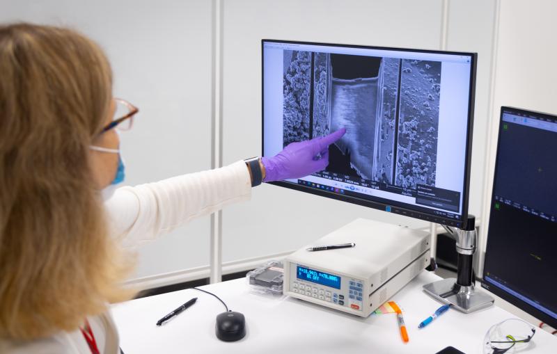
the invention of this camera so as I point out you need to record the images
now in the old day some of you as well as I am when you take a picture you bought a Kodak photographic female you
went to the darkroom and then you you develop the film that's what I used to do when I was a system professor I
actually make our own photographic film so that you become sensitive enough now
for this year they're actually electron detectors first with the core CCD camera
the other core direct detection camera and directly suction camera basically is
similar in physics that's why your iPhone but the only difference is these
are caused more than half million dollars a piece then the iPhone so that's only do and the physics are very
similar so now why is that useful and you turn out these dis camera that in
fact it was developed by the detector in in in in a lab similar to here the
Berkeley and lab in the Lawrence Berkeley Laboratory and so and subsequently pick up by by a
number industrial company that making it a commercial well available the the
scientific merit of this camera is at very high frame rates it turned out one
of the challenge in using electron is when the it's on hitting the specimen specimen will be moving so far if you
take a picture of a baby or me at this point then you will have a fuzzy pictures right so now but if your camera is very
high-speed then you can take multiple pictures of me and subsequent you can use a computer to align one frame to
another and so this is what this picture shows it you can actually align multiple frames in such way you will reduce all
this bearing effect due to motion of the specimen caused by the electrons now the
other scientific merit of this detector is it can come single electrons so that
means this extremely vision they though they waste no electron every year tron
coming in they actually can't it so they form useful images so because of these
you release the microscope turn out to be very done good for many many years
because the Optus opticals yet on microscopy develop those optics
were nicely but we were not able to record the images good enough until in
the last few years so i think these are the technological breakthrough i just want to point out what we get the camera
today is not reaching the limits of the physics set that means that's why we still need to do the research
then get the Poussin for the next generations of camera and so these that
means this view is the under active research so now so this that much technology i want to share with you now
i want to give you some real-life applications so for many years my dad
has been interested in a biological process the process involved is how the
virus are being put together in a living systems so it turned out the virus is
once you get one virus get into then you will multiple multiply many many virus
and that you you know the organism get in fact that would get sick so in what
part of the process is that the virus had the genome and the genome will be
replicated in the infected cell and then and then the replicated genome will
produce proteins and the protein will put it together in in a particles now
there has different color different color babies and different types of protein that make up this particular
cell or particles and then subsequently the DNA in this particular virus you
thread through one of the the the the vertex and then the gas boomerang inside
the chamber so if the outsides are all made of protein so so the water protein
the virus actually protect the genome so that makes sure the genome of the virus are not being true up by the host
enzymes okay so this is very important for the job if you have virus that you
have to have the right protein to encapsulate the right size it turned out interesting enough some viruses are big
some very small and for some for good
strategic reason of the virus the the chamber are the size of the chamber
inside the protein accepted the right side to hold the viral genome so it's me
it's a very precise process so when this is formed then where the entry point of
the of the genome will be C or by additional protein we cocktail and as we
saw these things sticking out would be become infectious can attach to the surface of the cell and then it will
then the genome will be released in the cell and then make the next rounds of replication so that's a whole process
now that tremendous genetics and biochemistry has been known in this process however there's no information
how this virus looks like okay as I pointed out in order to get the
structure you can do x-ray crystallography and in this particular virus copy 22 bacteriophage this
paternity to phase turn out to be a virus that can infer there's a marina now semolina some of
you may heard can be a food poisoning if you somehow go to some junkie pays to
eat the thing and you have food poisoning is salmon Anna so now how do you solve the problem I would sit there
maybe you use some of these phage okay because the face would kill the bacteria and then you'll be well again now that
is not a crazy idea because you think about you had about about bacterial
infection you know last thing you want to go to go is go to the hospital when you're sick because there are lots of
infection cap has to be done those are the bacteria so once your bacterial infection what do you do you would
actually have antibiotics right but sometimes antibiotic doesn't work so in
fact the Russian has suggest many many years ago maybe a face therapy you know
if you somehow feed the the patient with some face maybe you can kill the the bacteria rather than antibiotic now that
was I thought was crazy I did in many years ago but now maybe not too bad idea
because some of these antibiotics actually doesn't work but anyway so that's that's a Kinetico way of looking
at it but I'm interested because just the intellectual curiosity how this diagram are actually occurring ok so but
anyway these piton interface is the object that I will spend a few minutes with you how we can use the choir um to
a microscope to look at these three-dimensional structures and the last problem I thought we talked about
how do you look at the virus at these pages are multiply in the cell how do
they look like because so far in this diagram we biochemically purifier so first of all I can't like to share with
you how we you see yet on microscope to look at this mature of H P 22 so here is
using these expensive camera we take multiple frame and then after where we line up the frame now as you get more
frames all these this virus particles 70 nanometer big I become more and more
protons you can see that after 17 frames things become more statistically defined
so you can see these particles different from these this is all slightly different because
turnout this particle embedded in this matrix of eyes are oriented differently if you look at me this way would be
really different from that way so besides why this particle don't like look like this even though they are
identical wireless particle now so in our data collections this is some of the
old data we did some years ago so we collect about 20,000 particle in the
year time microscopes and then we combine computer and then we generate these structures and he structures it
actually look like we call buckyball and you turn our Bucky boy is was was
mentioned by curl quote oh and and smarty in Texas which I used to be in
Houston so they are or with frenemy they won the Nobel Prize in 1996 because they discovered these buckyball what the
buckyball said is these are only carbon atom in this simple case it has Penton
and hex on there 60 carbon in making of this buckyball now in biology in this
peteanddee virus it looks like exactly buckyball it also has hex on 1 2 3 4 5 6
and also happy pent on ok so he also have 20 set of these hex on material
press one pent on material so so I call these P 22
really a power to go Bucky ball okay in terms of geometry they follow very
precise geometry as the C 60 carbon as him as as discovered by professor
Smalley at Rice University so now what this diagram so2 is about the position
and if the density the atoms okay so I'm going to show you a movie in next line
to so use the spin around and I like to zoom in one of these 20 hex on in here
now the density are so good that I can use a computer graphic to color it the
- there's six of them their 60s in color each of these are represent a strings of
of amino acid to form a protein so there are six identical protein to form this
hexamer so I just need to say one word about the protein protein is a strings
of amino acids usually their small size of protein as a big size of protein in
this case is about 40 amino acid now in the metal I mean as a mega protein
there's only 20 different kinds Society bet we're 26 alphabet depends on what
what words you use and they're different combination the alphabet in the protein you have 20 alphabets and then it
depends on what combination of those amino acid alphabet to make one these
distinct proteins so now the the interest what we have in here is how
this alphabet amino acids are being string together in feed amount of space
it look like pretty nice good-looking spaghetti but they actually a very well
defined speaker it's not a random spaghetti in this string okay so now
then the question is how do I show you exactly how the amino acid in one end of
the molecule to the other end you know they don't the both end can be in any protein can be close together it can be
very separate apart so then there is what we call a modeling so we use a
procedure that we can actually complete a procedure can trace the we call
backbone this spaghetti polypeptide that I'm a no acid connectivity you know they from left to right top to
bottom and the top so you know this is a journey that the amino acid had put
together in order to form a functional proteins okay so it's not a random now
if your sake my Alzheimer's some of this these voting can fo exactly and that is
why these some of the protein can can get cause disease so by in this virus be
turning to that capsid protein form this particular trace now as I said this
movie show you the connectivity from one end you know this end terminal C terminal is on the other end so then in
addition to the connectivity you may ask why we are the atom you did I say the
the protein Amir atom so the next movie show you where the atoms are okay so now
we are now it is fret of amino acid we build the model with the model with this
y where the atoms are which we will probably be sending atom in different
color oxygen in red and nitrogen in in blue and then the white color are the
carbon because by and large the protein molecule are carbon oxygen nitrogen and
hydrogen okay and sometimes we have also a software as well so here give you a
model to describe the position of each of this atom so in this particular case
they are about several thousand maybe four or five thousands and a forty
thousand atoms or together so we are able to determine all these in three-dimensional space so now as I
pointed out these are made by axon and Penta Undine these in this fundamental
unit there 60 of that that make up the whole of paternity fate so now each of
these density we can build a a model and when we support those model it turn out
even though each of these protein is made up identically with the same chemical composition but it's but the position of
each atom are not exactly identical because each of the protein are surrounded by slightly different
chemical environments okay so so that is what we display here this seven
different color will be supporting posted these and of these you can frame out differently in each of these protein
in making up this axon and the Panther and this loopy are also slightly different which have expanded in a minute why
that's the case so that complete about our journey in from making the the virus
taking the pictures and currently concert in three-dimensional pictures
and then beauty model and and then so from here our interest is exactly what
are the interaction between the protein because remember that the proteins are
to protect make a shell to protect at the end our DNA so we discover that in
this model at the end of this n-terminal we'll be saying each color represents a
fan protease and protein so the question I was asking is how this protein being
held together it turn out it is tear in here the red protein and the brutal teen
are the protein between the adjacent axon they are being held together by the
hydrogen bond okay which is a very shortest and only a few enzymes so their
answer our first chemical questions how these six protein molecule to form this
hacksaw and it turned out in addition to some of the index and one of the great
in the accidents are these hydrogen bomb holding its side in other words one one
hand holding the other guys hands holding it together in a chemical sense so there is our first discovery how this
protein are being held in a stable way so that the DNA will sit comfortably inside the virus chamber now the next
question is how are what are the chemical interactions between the protein around the axon we are talking
so far this hydrogen bond is the protein across the axon how one protein from
axon to the protein of an other acts on the next movie I'm going to show you is how each of the protein within a hex on
being stabilized together so here I'm zooming in here again in a model and I'm
putting up the amino acid atoms as well I'm assuming in the interface between
one protein to its neighbor and when we examine it we discover that
they're about a different particular chemical interaction we call saw
breachers so that means these oxygen atoms in these amino acids are in there
with the nitrogen this but the special name for this harmony arginine and glutamic acid so the distances form a
hydrogen form a iearning interaction is a charge-charge interactions so what we
learned from here is about between the interface of the protein within the axon
they are charged and charged into actions now why this is important now if
you have a virus infections if you want every drug the drug what the drug can do
is can disrupt the assembly of the protein so if you have weight off of
disrupting these types of chemical interaction then the virus will be
falling apart and then you're well again right so that's a whole idea when you want to do structural analysis is to
discover what are the chemistry involved with the protein-protein interaction in
such a way that we can start thinking about a therapeutic intervention process
to disturb the virus so that we will be well again so so I think this give you a glimpse
how we go from images to map to model to
a chemical details now as I saw in the cartoon there's one corner of this
particle have some other proteins involved with the DNA encapsulation so
far I didn't show you that because these particular first by analysis I want to show you the detail of the cell the
buckyball cell protein cell how they organized and how they how they maintain the stability so if you kind of look at
that you know somehow there's something sticking out here as what the cartoon said okay so these are where noisy image
but we are able to actually the next step would be an other image analysis we
are able to give you a structures about this so I talked about the detail in here so this one I
can actually cut it open and then I begin to see their additional new protein they are one two three four four
protein inside of this capsid protein cell and I'm showing you each of these
like this blue protein has turned out to be six copy their crystal structures know about that I knew go put in their
band to get the model you fit into each of these six protein and this we called the middle of free protein and then on
top of it there's another I call purple protein and they turn out to be 12
copies and I go to put in a banked and fit that so I know this is purple protein and then again they make up the
same same chemical composition now I'm looking at this green protein I also turn out to be twelve copy a crystal
structure also long in each of these proteins and then now we spin around it look like what what a protein databank
looks I so as we saw we can now combining information on the cryolator
microscopy together with crystallography of some of the individual protein that
the cassava able to Chris I and sabi atomic structure so now we have a whole
atomic level description of this virus and in the last movie I saw you that
turned out to be twelve copy this portal green protein twelve copy they have pepper bobbing and acting copy of the
tails bite and then free copy of this middle protein or the GP is the name of the proteins according to the
geneticists and of course their DNA in their IDs they have time to show your DNA is turn out the DNA is form a poop
inside the chamber so that pretty much
finished what I want to tell you about these infectious paternity faders which
will infect a Sam Alana now so this is a mature phase but we are also interested
how would this virus look like before the DNA comes into the chamber of the
virus so then we again that means in these we call PO capsid now in these
as this cartoon say there are the blue protein which I went into a detail to talk about but they are also purple
protein and I also show you some of these other protein in this vertex so so
the next set experiment we did is to look at the structure of these capo capsid and so here is the experiment we
also took images in this expensive movie frame camera and this one I show you now
one thing you notice is these look like more angular the structure so it is by
more circular is not your your eyesight bearing there actually to you know the
one on the lab is more circular and you look at more carefully get a computer ruler this actually is a a smaller in
time there by around eighty Anselm's okay our eight nanometer smaller in
diameter so the interesting question is that means when the genome is packaged
in the virus the virus actually is swelling okay before the DNA is skinny
and run once the genome can in the angular and fact and the question is how
does it do it do they do chemistry or there's some mechanical motion so stay
tuned the next movie which I'm going to show you is about these structures on
this one before the DNA is encapsulated and this one I saw you in detail why go
and so now we did subject that the same they deposit saying you know twenty
thirty thousand baht ago and to process the data to get these particular density
map and then we then extract one of the protein in the next slide to show you
this is Sentinel is the same protein okay I color differently because this is
the PO capsid is that this protein that is the structure when the virus do not
have do not have the DNA this is the one I show you these purple color
is the model of the protein after the DNA's and capsule as you look at it you
know this long tail I can't flopping around in the mature virus whereas is hidden somewhere at the
back here and then you look at this loop reefing also slightly different and in a
technical term we say there are structural changes okay conformational
changes and the question is how they make these conformational change and this is a movie to which I'm off these
hacks on so this one is the mature wary on you can see these are a whole body
movement it's not just I'm not I'm waving both my arm and my foot and it's
all moving in such a way to accomplish these structural changes in the virus so
first in the mature very on this whole closing up again it's not surprising after the DNA come in the job of the
protein is to protect the DNA that's why they don't want they are open when you open the outside in the cell they are
very aggressive enzyme can chew up all the DNA there's no good right so there is a design recovery that once a protein
in there the protein and the conformational change protecting the genome and secondly the interface
between the protein also different so that means there's multiple conformation of variations before and after the DNA
is a camp capsule now here is a movie - - so - so in the side view you see this
enzyme and I actually dropping around a lot so a lots of protein move but they
some of the body move just a hinge motion but this one is definitely more
than just a hinge motion there's a lot of changes for the polypeptide of the protein so from there now I just want to
show you that in in in the virus before the DNA come in somehow the core
so is how they build the shell and the protein the cell does not form these 60
copies of these exons they actually need initially some protein to help we call
scaffolding protein so these we looking from outside and then you use the computer to cut it and to say watermelon
to look inside their red protein now I just cut a red they are not red okay so
so these turn out these density red protein because scaffolding protein and they are kind of one to one ratio one
scaffolding protein by one of the capsid proteins they have the cell to peel so that means in this building process to
get the the the whole whole virus into a stable buckyball before the DNA comes in
you need assistance of this red protein cause covered in protein and now as you
saw the movie that when the day they come in some time is moving the reason
is moving because some of these red protein has to get out right because there's no space when the DNA comes in
discovering protein I get out to let the DNA a and that's why that last terminal
portion that you see the movie moving around they're moving pompe actually was
bang to this red protein in in the pod capsule form but once it's mature then
the DNA had to be released from discovering protein undergo this
swelling to make it a tea and some wider capsules so this conclude my
presentations of how this virus after in two different conformational States
after the DNA come in becomes infectious I show you the structure for all the
protein power and also so you or the protein part before the DNA comes in now
all we're done because we are able to purify these each of these separately in
the biochemistry laboratory now what happened is a well why you are this and
in the ivory tower you do things in the test tips are really is
I raise the same question to myself right my daughters are there what are you doing you know how are useful your
thing doing how's it oh it's going to be useful one of these days so now I'm showing you now let's look at the hotel
right now I'm looking at the hotel which have been infected by the virus is that
really enough I think it's really enough so now but the cell as I saw you in this
scale diagram they are about hundred times bigger than the virus that means the the bacterial cell can hold tens of
this this virus particle okay in addition to all the protein machinery in the bacteria so now when I look at this
I turn out this particular cell is a bacterial cell in the ocean because
cyanobacteria now why is interesting because in it turn out a whole pile
ecological system we want to come out the Sun the Sun give us the energy the
energy will convert into organic mass and so channel in the oceans the flow of
shock you know a lot of fish and shark but in addition there are lots of bacteria swimming and Iran as well this
poor bacteria they may be eaten up by the shock but anyway I don't know that so but this particular bacteria is
useful from the point of view to understand what the most fundamental process in life is to capture the
sunlight to make organic mass okay and number one and number two each of the
bacteria in our cell in in the ocean they are also associated with features
okay that is in other words these phases you know if all the features die the
bacteria probably die also it all the bacteria die eternal in between us a lot
of bacteria hopefully we have all the other defense mechanism to make sure that the bacteria is not too offensive
so there's a kind of homeostasis or right now in the medical school that you
know their cancer patient eternal you the cancer patient some of the complication is some of the bacteria do
they come microbiome it's not imbalance okay eventually one may die because of
these bacteria come come mess it up in terms of things so now when I look at
this this is about fractions of my card they're very thick okay so you see this
one what are you suing it's just a black thing you know I pay you my text dollar
is not with it so I said don't worry I probably can do some physics which I'm
macaron with these putting a face space in such a way I get these compared look
at these and then from nothing to something I'm not praying magic but this
real there's a real physics okay so that means that's why we need physicists right because they do good things
sometimes most of the time and so so so
we call a faceb a meter so Nikki you also got the Nobel Prize a long time ago in the light optics invented peace but
we applied it here to an optics and so as a result of that then we can look at
these by we caught tomography it turn out every bacteria is different from any
other bacteria so no two bacteria identical so that means we need to get a 3d pictures of one back here at a time
so the way we do it is in the microscope we can actually rotate the specimen
looking at different angle so that you know we can get multiple pictures and then just by a CT scan we get a tomogram
we construct it and here is lie in the microscope we can tu this bacteria and
then again before data processing you don't see much but now the next movie
I'm going to show you a tomographic reconstruction of the cyanobacteria infected by the sign of ages now you can
see there are billions of protein molecules in here you can see all kinds
of features in a year this is the outline of these cyanobacteria all these
are different protein machine this wrong thing right here I'm zooming in at the
height on these reconstructions and so again these are very congested informations I'm now showing you a
different representation of this cyanobacteria I'm flipping Iran in the
tomogram in a computer after the reconstructions and then subsequently I
use a a computer color pencils by hand these other we call the cell membranes
and then because of Farrakhan membrane is the photosynthetic reaction reactions
here there's these silent color things containing the the enzyme that fixed
carbon dioxide in photosynthesis reaction these are infecting features and finally
you see a lots of purple thing these are newly born features muley since I've
synthesized features that we were able to capture in these tomogram so now you
can imagine what I show you why I color it is only a few of the highlight you
look at these these phases actually drill a hole into this membrane to
transfer its DNA into it into the cell so we to see a difference light leaking
because there's too many features in their suicide a leaking some of the other things are going outside so as you
can see the one I said color pencil I really meant that you said computer so
the color you saw it took us a week to Korea now fortunately I have very
diligent postdoc who was willing to do it but after he/she did once that wha
I'm tired I said what do you mean tire work harder he said no I'm politically incorrect I
should encouraging loving care to my student and poza so I said okay so in
other words maybe we don't need to work harder maybe we were smarter so in fact
another student come by they said why you don't need to stay there for one month you know to tolerate as a kid and
began graduate student and I'm from Stanford I must be smart right so then
well I was from Texas so the stacks and also smart to in different way and so so
we eventually employed these this album called deep learning okay deep learning
is the artificial intelligence so we actually develop in such a way all the
things we color we can do the research computer using in one-hour time instead
one week human time so it's really exciting you know when we first did this was really labor-intensive but now you
know you can watch a football at the same time that the computer color for you I still intend to do that
you know there's intelligent people are Stanford so now in addition to this I
said okay let's be real we are not only looking at the bacteria infected by the
phages only in one time point we need to do multiple time point you know right you get in fact about
active bacteria usually your temperatures okay such a high temperature you know because the phages are multiplying in you so so this is
experiment we did and different time point it kind of grooving the different
time point so at the beginning there are no features in there when you in fact there's some features in the outside and
then at some stages there's some inside and outside at the end they're print their newly synthesized features inside
so we can actually start sampling different data point equivalent to a
different physiological steps okay now what I'm saying here we cannot synchronize these in the infection but
getting a lot of data we can dr the example to so that so then in here then
then all the purple beads here i said they all look alike I said maybe not let's look closer let's
put on a many fine gases computer Marin fire gases look further and again thanks
very much for the image data processing step in such a way that we actually was able in those tomogram to discover their
five types of particle they look different so one knock roundish and
there's something inside the they got cutaway view this is the surface view and then some you see
something in one vertex some you look angular and have something inside some
si tail and in this particular sino features eternal we call a horn on the
opposite end so in other words in this vast amounts of tomogram using a
computer sorting a lying and everything we are able to discover five particles
of different size and shape that's real right they turn out in this project which is different from p22 pretend it
is infecting some antennae disinfecting this I know bacteria has this horn
one of the question among these face biologies when does the horn get on
touch to these rages by just look we have this assembly the horn must be
assembled to last so as we saw we are able to figure out the pathway how this
being assembled ok now interesting in app this you actually see some density in here I said
that the DNA has to thread into the into the chamber of the phages it's no female
it takes energy it turned out this is an enzyme provide the energy to package the
DNA in there now you also turn now in these particular features we are able to only purify this we were not able to
purify or the other easily so what it tell me is if I want
to look at the structure of a biological process in this case is the virus assembly is a is a in fact in a sensing
infection process of a pathogens if we understand how this assemble in this
process we can again start thinking about what drug we want to use to
disrupt either at this stage to go from this or don't put up the horn at the end
ok now this only back here only the cyanobacteria we don't meet the drug boy but if you can imagine is a herpes virus
or the Zika virus which has similar types of buckyball arrangement of the
protein we can think about a drug design or an antibody to neutralize these kind of
pathogens so as a result of that we can actually work out the entire pathway of
the SMB from the PO captured with the scaffolding protein the the DNA and then
in fact you know in this in in this one here this is more a diameter and this so
these also so that the swelling of the capsid happened at the very onset of the
DNA packaging then one of the question when does this get swollen into 70
nanometer wider it turn out wonder DNA comes in is that swollen not all the DNA
come in and swollen so that's again I can't mechanistic process we were able to to delineate through this kind of
thing now so some of these process could work out by genetic manipulations but
those are very tedious so again my mantra is just look in the nature
everything you just mean having powerful yet a microscope a good computer good
software hi lots of expensive survey guy do we have a problem solve and so
finally I can't decide to summarize the opportunity for choir um research in
biomedical sciences so what I show you is a phages is a bacteria but the same
technology in my lab and many others can now apply to multiple team molecular
machines that have relevant to diseases so I saw you in the first two example we
can actually solve the atomic structure of a molecule machine in this case ages and which the phages can have different
conformation in different physiological states like the POE capsule before the DNA comes in and the battery of ages
other than they come in the structures are different we can do it without involving crystallography in fact these
phases have been tried by Chris father for many many years he was able to crystallize too soft
structure so that's another why you on doing quiet an alternative procedure other than
Chris of they give you atomic resolutions structures now the other one which I am patella interests looking to
at the future is to get an atomic resolution structure would he need to enable for drug design and enzyme
engineering nanoparticle and synthetic chemistry now all these big words but
that's for you today we are sake we need drug okay a lot of drug company are making drug before you make drawn you
need to know the target right in this case a virus and you need the structures
once you need the structure then you can in tight anything about where are the chemistry holding the protein together
in the in the protein machines and in such a way with the vector so now in
these technology is now fast enough that you can be part of the pipeline in an
academic environment or in industry of the farmer to use this this this as a
data bond for a drug discovery process and nasi then what I'm excited about is
we can directly visualize the maraca machine by the phages in situ in the
cell okay in the cell either in a normal state and and pathological state I
personally now we are project in Maui Huntington disease which under the disease I involve in the protein
misfolding so what we are interested is if if the individual hypnosis is how
would the cell reply how they look different from the individual term health or diseases so I think by
understand the underlying organizes a social organization we hope to find
solution and therapies or or prevention
medicines or to to overcome two diseases I have two most like to make you think
big I think to convince you that actually drug this I is visible now in
terms of this I what I saw you is only three and half three point free end zone
the medicinal camera say wow maybe that's not good enough
because I really need to get in crystal clear each of these oxygen nitrogen atom
and can you go beyond three and a half and my answer is yes because you pay me
well so here I came to Stanford we set up a microscope this is called April
ferritin okay April 13 is another molecular machine to hold up all the iron you know some of you need iron
every day you know this is the peel right because they are actually this April ferritin can contain the eye and
the supply to the body so these are the images of these April 13 and we solve
the structure look like another nano cage now I color it four different color
this time it turned out instead of 420 protein in this case only 24 proteins so
each color is one per peptide and in fact if I extract that be a model only
one portions of Puerta we actually see really detail live for example this
amino acid okay right here we call a ring structures at one point I handsome
this aromatic residue look like doughnut doughnut at a hole in there if a
low-resolution look like pancake pancake does a Navajo so I think these kinds of
information is what the medicinal chemists will I at the farmer looks like so the good news is the quail yam can
actually generate these quite rapidly and dr. climbing stem in in our group
were able to collect data of the soil in ten hours time to January in up data to
create these kinds of density so I think the good new is this is that we finally come in the place that means the
Stockholm Nobel Committee did it right this time it is actually mature not just
for academic interests have a translational value as well now one more last one about looking in
the cell which I'm extremely passionate about is I work when I was in Texas working
we profess a a new soap who's a ovarian cancer oncologist so based on his
studies there are wearing cancer patients the prey that is from the blood
tend to be somewhat abnormal this the patient seemed it from both eggs so we I
said there may be we just look at it okay so here are this first clinical research so the clinical research I did
actually from a patient sample so these are the normal subject in other mega
sort the interesting part are this blue protein called microtubule they can't
wrap around the play that in the normal individual the Pinay patient these steve
wrapping around but study broken up but in the Pinay patient they're all these micro musingly broken up okay
the interesting aspect about these observations is that if this quiets on
tomography method can potentially be a tonic procedure in my opinion okay so it
turn out or wearing cancer the same any cancer is very hard to know until you
really at the terminal stage so my idea is why every healthy individual just
carry a job abroad every year go to the doctor and then put in my microscope
will tell you how good you play that looks i now if you somehow go something wrong with just one year at the time so
i think potentially it can be a diagnostic purpose and it's interesting
enough according to doctors Oh in the ovarian cancer patient the diagnoses are
not very good with the with the current diagnostic procedure so just like many
cancers like prostate cancer before PSA it was good but now we were told maybe not that precise so I think the medicine
is still a evolving branch of of sciences so I think I personally feel
quite yet on tomography may potentially have a role to put it in the clinics and
finally and because all these excitement and Stanford University and slag decided
we need to do in a right way to set up these kinds of research scenario and
food a very generous support from the president of Stanford and the director of slack and the Dean of research and
many faculty we set up a query emphasis it is only five minutes walk from here
and so we have all kinds of stay about instrument and we have for high-end
instrument we as freezing appraiser specimen preparations in the building six I really invite you some of you in
the future the semi email you're always welcome to come to visit us to take a
look how we operate in in in regular hours I just want to point out why we
are talking some of the people sitting at the back in my life probably collecting data in other words some of
these data collection can be done remotely and so in fact some of the
project the post office really now become a professor now hung row and he
collect a data set on one of the channel in Friday why he was attending a conference elsewhere and there was a
sensational data he got so now with all these investment
from Stanford and party from the National Institute of Health the necessary set decided they wanted to not
only people at Stanford can use it he the NIH through your tax dollar support
decided that we wanted this facility open to anywhere in America so they pick
up three sites and we are one of the three sites to set up a national cryo-em
Center and so we are going to open the center's up we just get the funding
about two months ago and we are very excited that we will double our capacity
not only for our own local use as Stanford and slack we also actually
welcome anyone in America and Beyond to come to use the facility that we're
going be really exciting there will be a lot of biological material weekend first
time to look at the structure and function hopefully it has a
translational value to overcome to combat diseases and also solve the the
the Eternity by energy problems and finally all the world I see sends many
capable individual when I was in Texas some of them actually move along with me
that individual and the face project was in long-term collaboration with
Professor Jonathan King MIT MIT just don't have these kinds of things right
so we have been working with him for many years so we are really excited with
all these colleagues in my group who contribute a lot of whether they
dedicate time for many many months and year to the story I present to you I thank them and I thank all of you to be
here tonight thank you [Applause]
thank you you'll take questions yes so we are
taping this show so if you have a question please raise your hand and then use the microphone in front of you
there's a button for that press that questions yes yeah you can just use the
button is pressed one what is the function of the horn on the virus okay
what is the function of the hole in the virus in the POE capsid it's as a whole
because as I mentioned there's scaffolding protein in the POE capsule
somehow the scaffolding has to get out now some of the people get out right now they go to here and but the view and get
out you need to go out a way because the DNA go into one entry you cannot get out that way so the whole allowed the
scaffold in putting the get out excellent question hi so you mentioned that it takes about
10 hours per sample is that all the way from the protein biochemistry to the 3d structure or is that just the
application so this case is extremely lucky case that's like anything you know
some of the project we need even days and months we still don't get close to
it so this particle a preferred answer now is a well behaved protein machine so
after we purified then we we freeze it you know purified probably take X hour
more than 10 hours and then freezing it is pretty quick is 30 minutes and then
in this case we only use this automatic data collection in 10 hours we were able
to get this and in fact if we do only one hours if we're lazy then we get to
point for enzymes no free meal so you need to work harder to get better resolution
so have a question about water so you have oxygen in your image how do you subtract out the water background and
then also does when you freeze it does the water bonding affect the structure and alter what you see very good
questions okay it's when you freeze it when the water is frozen you turn our water can
have different phase transitions it can be crystalline water it can be amorphous water you know the water molecule x 2o
can forming crystals so the way we did it octopus a who got the Nobel Prize was
because he was able to fix it very quickly in a very efficient
quieten called a thin liquid ethane so when we first did it at Berkeley with my
mentor professor Bob Glazer we foresee in eco nitrogen it turned out in liquid nitrogen it was not frozen fast enough
so the water become a crystalline ice and then Socktober say change that
method using a fan and that you can close very very quickly and the speed is
something like 10 million degree per second that quake and so the water will
be in a Morpheus Icefall okay and that's how to keep the specimen hydrated and
MPSF well did I answer your questions the other part of it was how do you keep
the oxygen but even from amorphous from showing up in your image is it just it's random no oh okay so one of the thing is
you may be asking do we see water in our map okay I didn't have a sigh and in
fact we did at the one point I answer we see water molecule and the question is
how good the resolution you can see what a molecule you know I explained if you have something like two and a half and
some resolution you begin to see the water now what is so little then one other thing I didn't tell you is how can
you trust me what I said okay after all I came from Texas right so if
they were good stay so I think this is a good question so that our community in a
choir um community we are very concerned how do we think what we see is what what
actually is so we we undergo a lot of validation procedures they're
computational procedures to allow us to validate what we see is actually what we
mean and so so our experience that - so
one of the way that a couple of structure we did they also accompany
x-ray crystal structure they also saw water and and x-ray is
such crystallography is such a well established procedures so even though
some of them are not quite right as well but but in this case if they see the water we see the water at least we are
converging we could be both wrong in the absolute sense but then we need to interpret in terms of the chemistry one
thing I did not talk about which other example if you have an enzyme reactions
okay which William R water often time and whether the water at the right place
during the enzymatic reaction that's important so in addition to a very
rigorous validation based on the structure procedure we also need to make
sense in terms functions and then and then other thing we do is also we can do
mutagenesis we can modify certain residue in such a way water won't get
close to it and we can do another structure and then that that water molecule or whatever ligand and in fact
we also one of the pods on an ion channel we begin to see not only water
but we can see the fat the lipid in
complex as well good I want a very
exciting work how important is the knowledge of your protein sequence to
get these sub 2 angstrom structures okay so most of the projects we do we are
kind know what are the chemical compositions and and most of them
actually have a gene sequence and also compare a company with the protein sequence so in terms of the building
block amino acids we can't know in most cases but the question is do we really
know everything we see in there the answer you we already come across two
cases we actually discover something we didn't know before okay so in one of the
case we thought is only one protein it turned out there's another protein a small protein escaped escaped the
knowledge of of the biochemist who fought is only one protein when we finish all the structure and we build a
model we build the model according to what we were told on amino acid in the protein and we thought oh there's
another density I won our amino acid what's that it's a no it's another protein and they trigger us to do
another biochemistry experiments or another genetic experiment to confirm what we suspect another protein indeed
is another protein we are they come across two cases but it's an expensive way of discovering another protein but
that's okay it's very powerful techniques
thank you for the very nice presentation I have a question so during a presentation
luckily yokai Liam structure actually matched with the x-ray crystallography Oh x-ray structure so what do you do
when those two structures don't match oh good question in fact sometime they don't match and
then who's right and I would say all of us a right for different reasons because
it's correct because in the x-ray case they're very rigorous right and they get
one point five handsome they see every atom very clearly but in the x-ray case you need to crystallize the protein so
that means you need the protein in a different chemical environment by crystallizing it you cannot just
embedding in eyes you need to put some other chemical which chemicals don't
exist in the cell but they are doing chemistry but we are doing biochemistry
so we are interested by ofing okay so that's what we are after the truth the
ground truth in the native structures so we embedded in the chemical environment it would be mimicked in the cell so as a
result of that now the kristov I actually get interested to the cryo-em because what the structure they see you
know they see how the spaghetti are being folded in 3d those who had to be right okay but in the canticle
environment that we are in the cell or in solution they may be different so
that's why now usually the cassava also begin to do require yam to keep them the
structure put that in context
sorry to follow up on that um is there a third technique to where if I call him
because it sounded like if the if those two structures do not match the current
convention is now to just trust the Hyoyeon structure yeah that social media
see attended to that which I have no problem but but on the other hand I
already confessed that in our community we have to be self-disciplined that
means we have to establish very rigorous policies and procedures okay you know I saw you all these colors
signing thing you know we always mislead by all these color good-looking thing but they can be wrong I mean in fact
some of the choir um structure in the nature and not entirely right we define
any science it's not they ochita or anything it's just that if they have not
followed a very rigorous procedure they may be over you interpret on their data
and we did see that just like any fear of Sciences with the suggestion of using
the imagery for for Pharma is there a way to know like what the salt bridge is that you're also not attacking healthy
proteins that may have the same mechanism yes yes you can go either way
you can mess it up salt breaches but you can build new so pitches you can go either way
depends on the circumstance are you from the farmer good
we'd be happy to talk to you hi I actually have a series of questions it's
not that many three if you wouldn't mind engaging in a small conversation how
long is the wait list for the users to gain access to these microscopes oh you
mean here locally yes through your facility well we actually have developing a more with the new center we
have from NIH we will we will organize in the same way as the Synchron x-ray
beam nie that individual investigator I need to propose make a proposal and we
have an independent expert to give re the proposal not me so be to be fair and
unbiased and then they will score the proposal and then we see it's only 24
hours a day right so an expert that need to be exact why hours and then so we
allocate that and depends on the merits of the projects and then they will run
accordingly so that is what we are going to do but right now with the instrument
we have so we have Kearney of sufficient
resources to accommodate the need of the people on campus and slack and I run a
couple of NIH Center which I have to take a last constituents life excited
just finished writing a report we have the operation was started in January of
this year in seven months time we have 32 principal investigator professor from
different part of the world and we have solved 20 new structures how are these
32 and so nobody feel unhappy yet
so I actually try to accommodate as many people I mean in fact we are running the
instrument 24/7 it's nonstop the reason I ask is for the following
two questions which is based on the data the amount of output that you have given the four microscopes that you have and
running them 24 hours a day it sounds like one of the major blockers and being able to produce is the availability or
the time it takes to produce an image so my question to you is do you have any vision or insight into ways to improve
the time it takes to produce good questions so I think that there are two
ways that multiple way of doing it one is the increase of throughput so one way
if you have a camera that can collect the data faster is one way another you
can manipulate the beam make sure the beams do you could Cordy but the same time get couldcould images and the third
way is I need to pass the head if you have any money that will probably the
quickest way so so there are multiple ways so be sure to tell your friends to
recommend you for the nobel price when you come up with those improvements okay and the last question is where do you
see this technology or what you're currently doing going what's the vision
for the next okay so I think is going to be why not only those people you know
the biophysicists doing these you may want to ask what are this what are the limiting factor to use the technology
you tell now is to making the bhutanese material yeah what I show you this virus
is because professor Jonathan King you know he was very good in making purified
this virus seemed a perfect virus for this technology I mean we work on this project for 30 years before we start
means about 950 and some resolution when we first started now did we answer it's
a long way but he's far it's always good but our technology's not quite there yet so I think what I think the challenges
is the biology had to learn how to make this specimen so that we can put in the
microscope light this afternoon there's a pathology from the medicals who came
to to me and he was interested in a in a protein that has to do with hepatitis
virus that caused liver cancer disease and so and the questions I have is can
you can he make good protein so that we can do this guy high-resolution well so
I think that can be a challenge for some of these but I would say there should not be a fundamental limitation with
just nobody yet work on the biochemistry of their particular protein again if we put the right people that can be solved
so so I'm very optimistic there are chemical ways in making the protein to
be amenable for these methodologies so I
can see this technology will be a regular date upon on any power logical
research because later synthetic biology you want to synthesize new protein to do
new things new protein machine to new things we need to characterize it and in fact this cryogenic yet so my kospi not
only useful for biological sample it can be useful for non biological sample like
beam sensitive materials for example one of the professor in the on campus is
interesting alternative material to make battery how can you make battery cheaper
and last longer and so so but turn out those material where beam sensitive so
again we can use these cryogenic yet on microscopy to to look at those material
to aid the design of a perhaps a better and cheaper and last longer battery
material so I can see these methodologies not only for basic
research that I'm talking about could be material science research as well in the
medical school I think you know it's just you think about how many medical school in America maybe 100 maybe so I
don't think there are yet only a handful of medical school have this kind facility so I can imagine that would be
come embrace how many Hospital in America there many right if my is my
prediction is correct can be a can be a hit on a stick thing you know just like
a CT scan they expensive nobody asked me how much it costs in each days it's very expensive
you know it's cost more than a house in Perito so maybe around in the a certain
area house by for microscope
[Applause]
[Music]