SLAC topics

Cryo-EM RSS feed

Cryo-EM allows scientists to make detailed 3D images of DNA, RNA, proteins, viruses, cells and the tiny molecular machines within the cell, revealing how they change shape and interact in complex ways while carrying out life’s functions.

Related links:   
Joint institutes and centers  
Cryo-EM fact sheet (pdf) 
Stanford-SLAC Cryo-Electron Microscopy website
Research associate Megan Mayer and graduate student Patrick Mitchell load a sample into a cryogenic electron microscope at SLAC.

News Feature

In two new papers, researchers used X-ray crystallography and cryogenic electron microscopy to reveal new details of the structure and function of molecular assembly...

A model of the Lsd14 molecule
News Feature

Their work helps reveal the inner workings of cells and the behavior of matter under extreme pressures and temperatures.

News Feature

SLAC and Stanford scientists used it to zoom in on an iconic RNA catalyst and a piece of viral RNA that’s a potential target...

A high-res 3D ribbon diagram showing the structure of part of an RNA molecule
News Feature

From the invisible world of elementary particles to the mysteries of the cosmos, recipients of this prestigious award for early career scientists explore nature...

Panofsky fellows
News Feature

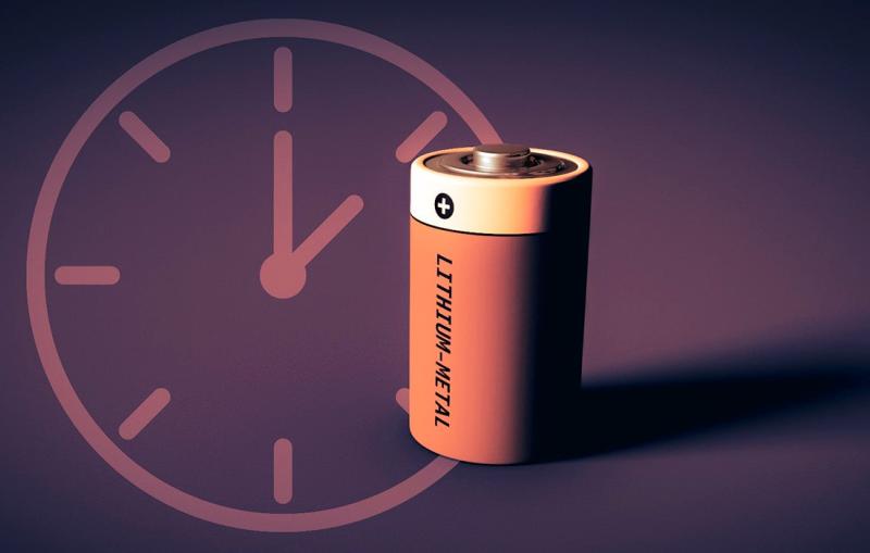
Scientists have documented a process that makes these next-gen batteries lose charge – and eventually some of their capacity for storing energy – even...

News Feature

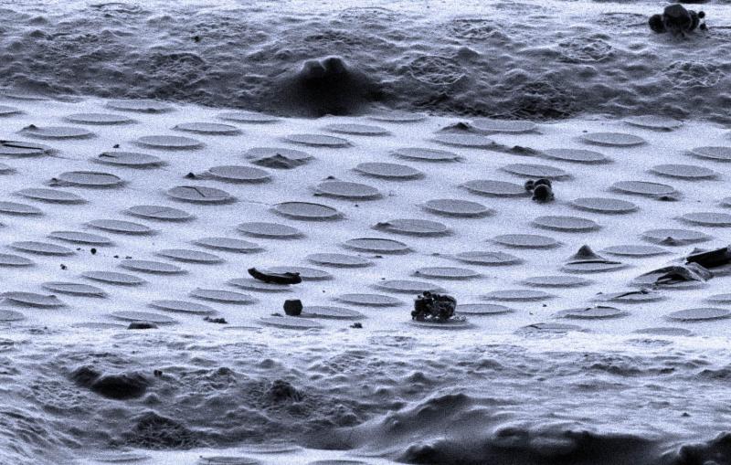
Stanford EM-X brings hundreds of researchers around the world together to discuss the latest methods and discoveries from electron microscopes.

Black and white electron microscope images of pollen.
News Feature

G6PD deficiency affects about 400M people worldwide and can pose serious health risks. Uncovering the causes of the most severe cases could finally lead...

G6PD enzyme in red blood cell
News Feature

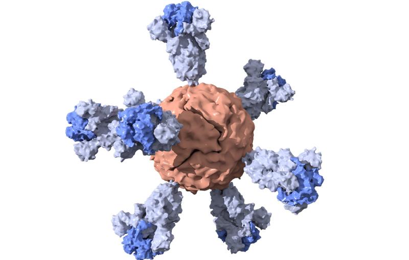
Researchers at Stanford are working to develop a single-dose vaccine for SARS-CoV-2 that could potentially be stored at room temperature.

The ferritin nanoparticle, shown with red center and six blue spikes.
News Feature

The study, done on a mild-mannered relative of the virus that causes COVID-19, paves the way for seeing more clearly how spike proteins initiate...

Illustration of a coronavirus spike
News Feature

The lab’s X-ray laser recently joined other facilities in making remote science possible from any corner of the world, a trend that will likely...

Remote experiments in the control room at LCLS
News Brief

The center complements other NIH centers at SLAC and elsewhere that broaden access to this cutting-edge technology for biomedical research.

Cryo-EM images of yeast cells with contents highlighted
News Brief

Images reveal how some antibodies may block SARS-CoV-2 infection.

A rendering of the SARS-CoV-2 virus.