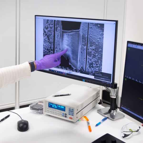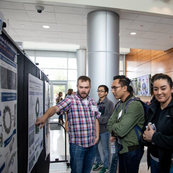
Photograph
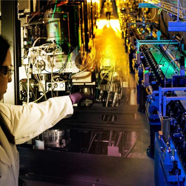
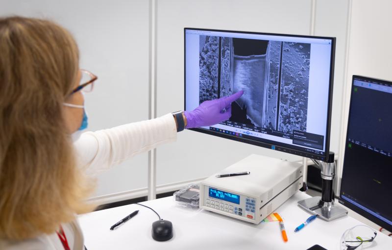
Lydia-Marie Joubert is pointing at the result of laminating an organic sample down to 100-300nm thickness for cryo-EM imaging. For...
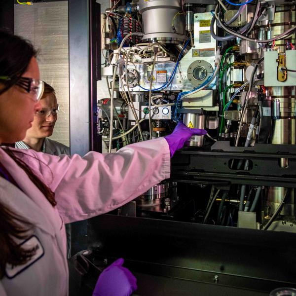
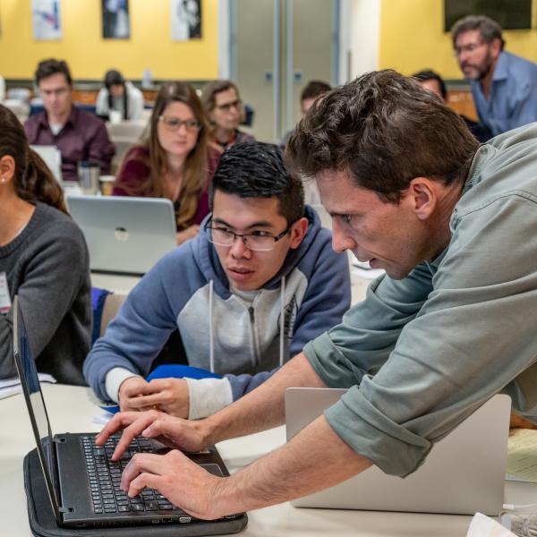
Photograph
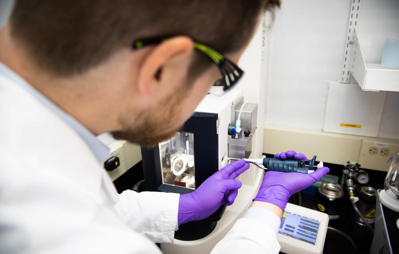
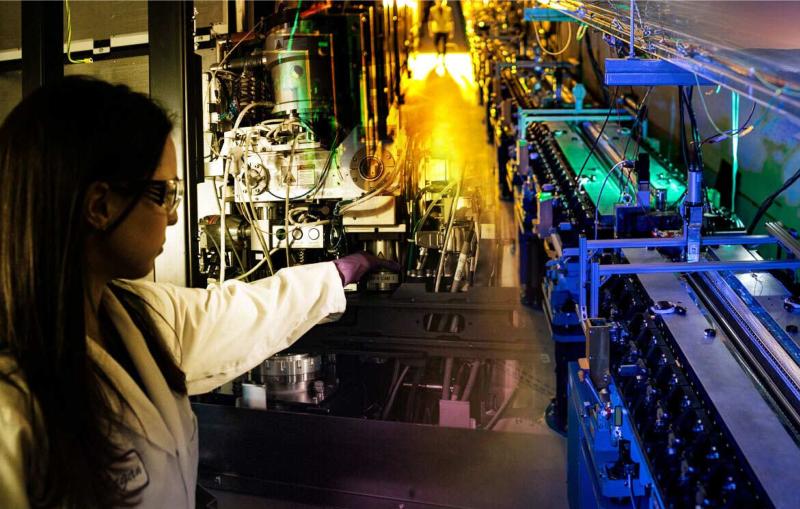
Research associate Megan Mayer and graduate student Patrick Mitchell load a sample into a cryogenic electron microscope at SLAC.

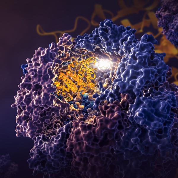
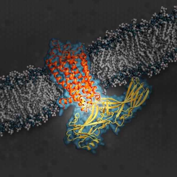
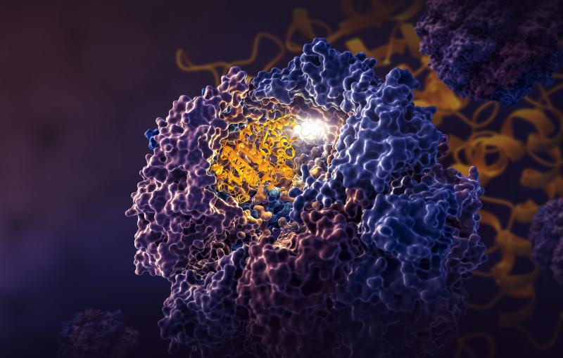
Illustration
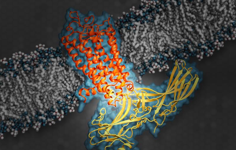
This illustration shows arrestin, an important type of signaling protein, while docked with rhodopsin, a G protein-coupled receptor.