Feature
VIA Stanford News
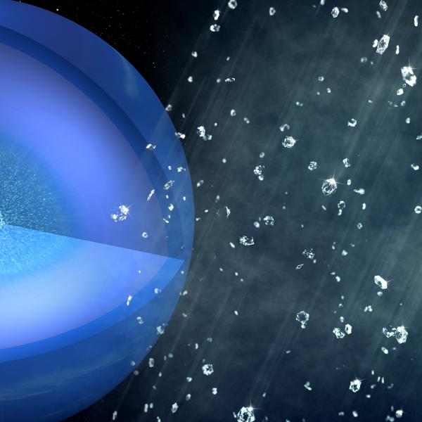
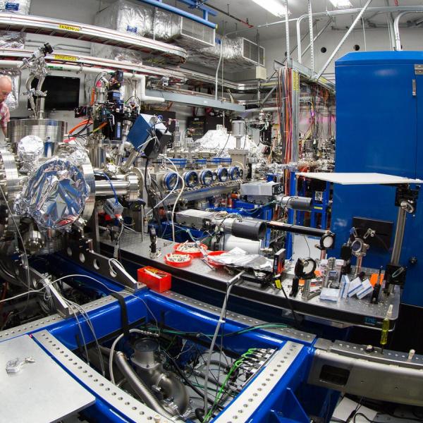
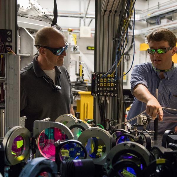
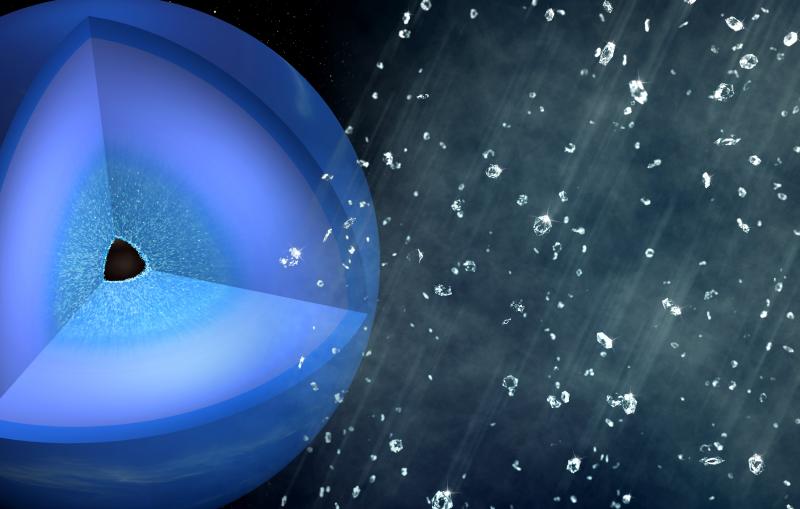
Scientists Discover How Dense, Extraterrestrial Ice can Form in Just Billionths of a Second

Feature
VIA Stanford News
Shunned by Microbes, Organic Carbon Can Resist Breakdown in Underground Environments