SLAC topics

X-ray imaging RSS feed

See content related to X-ray imaging here below.

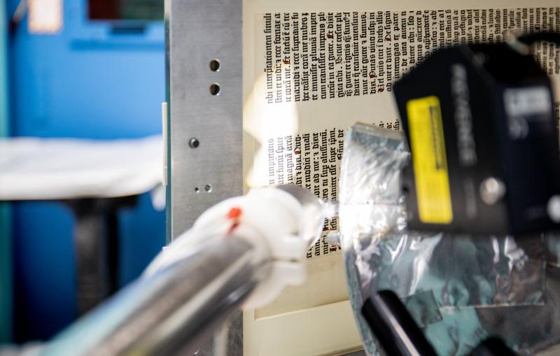
A page of the Gutenberg Bible from 1450-1455 AD is prepped before being scanned at SSRL beamline 7-2.

Press Release

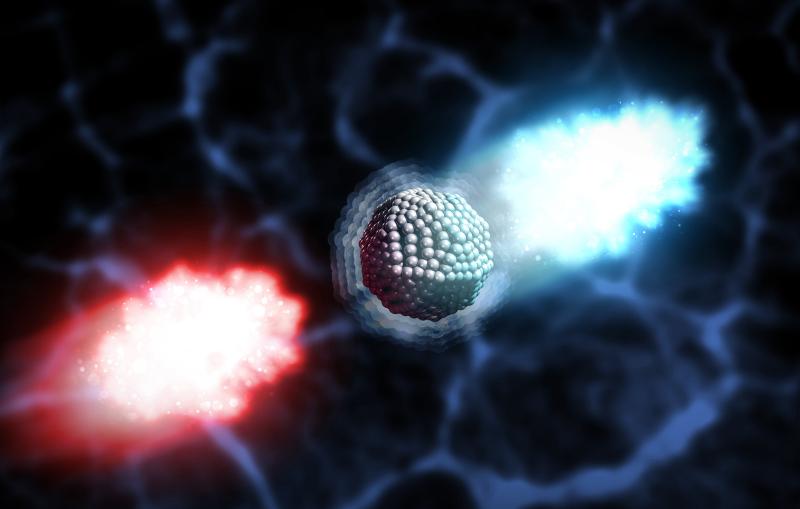
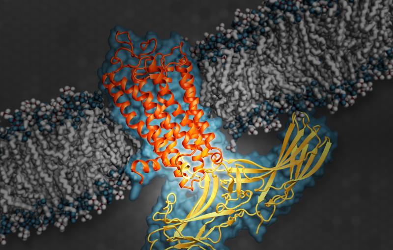
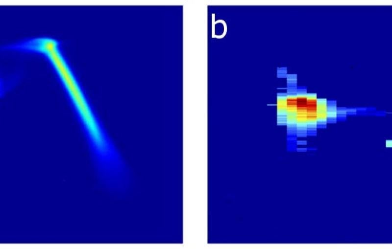
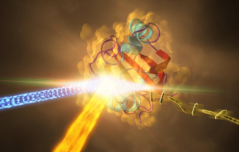
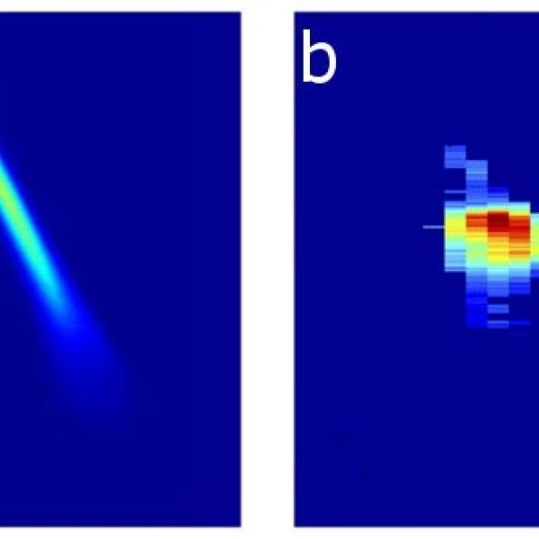
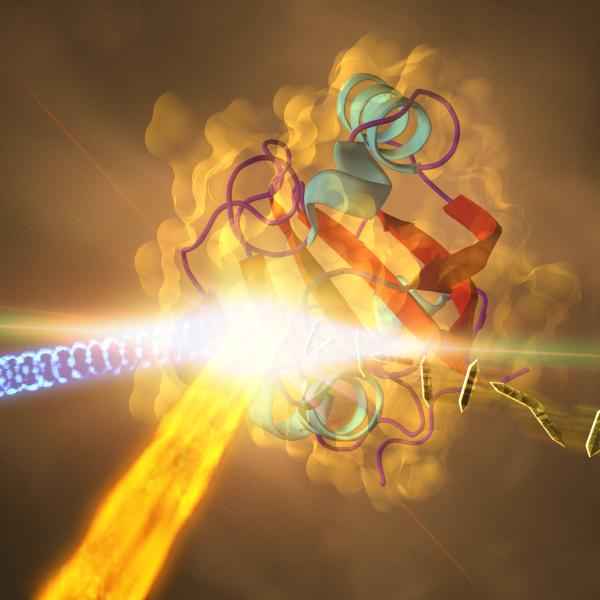
A new study with the LCLS X-ray laser could change the way researchers take atomic-level snapshots of important biological machineries, potentially affecting research in...

News Feature

This surprising finding has potentially broad implications, from X-ray imaging of single particles to fusion research.

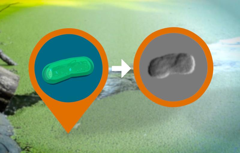
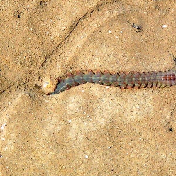
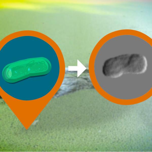
X-ray research on 80-million-year-old fossilized burrows, likely the work of tiny marine worms, is helping scientists understand how living organisms affected the chemistry of...

Image - This marine worm, commonly known as a ragworm, can grow up to 4 inches in length. It is part of a class of worms known as polychaetes. A far smaller variety of polychaetes was likely responsible for creating ancient burrows studied at SLAC.
Press Release

A biomedical breakthrough reveals never-before-seen details of the human body’s cellular switchboard that regulates sensory and hormonal responses.

 Illustration shows arrestin (yellow), an important type of signaling protein, while docked with rhodopsin (orange).
News Feature

Anne Sakdinawat, a SLAC scientist, has been selected to receive a grant to advance her work in producing and using new types of X-ray...

Image - Anne Sakdinawat (SLAC National Accelerator Laboratory)
News Feature

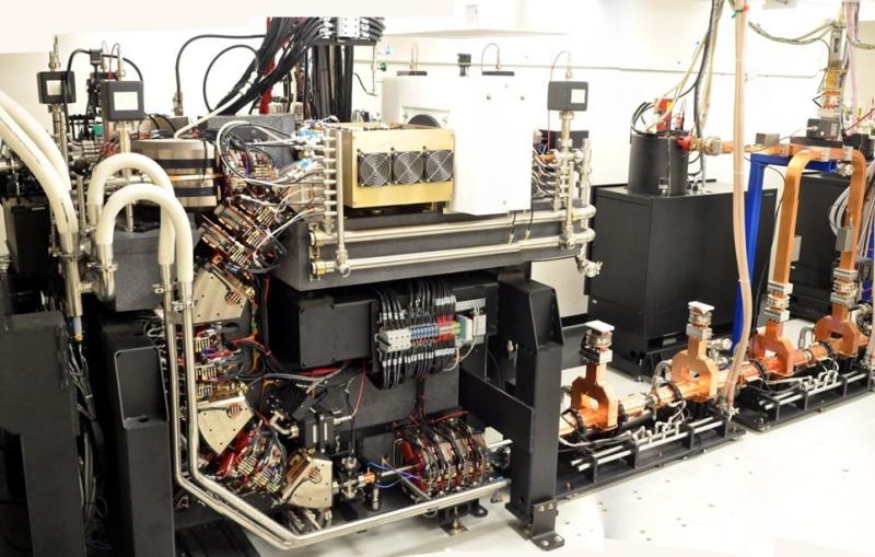
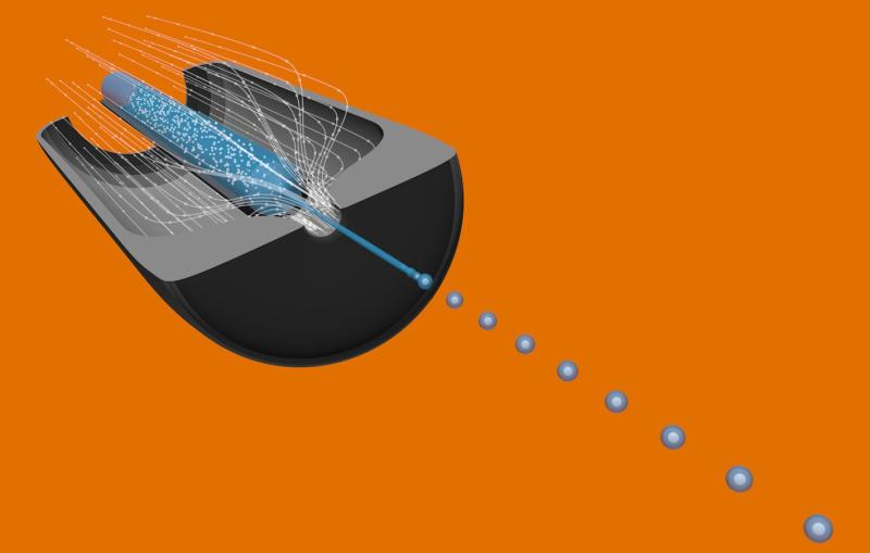
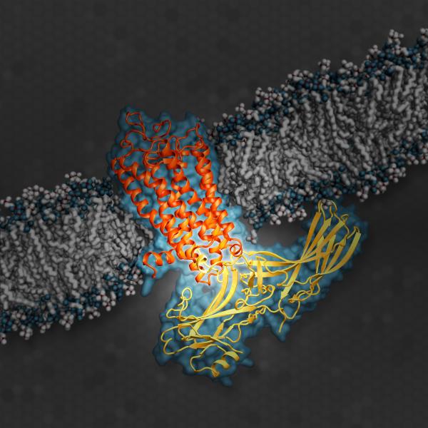
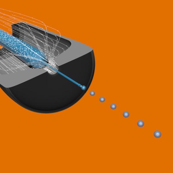
A commercial X-ray source with roots in SLAC research enables multi-mode computer tomography scans that outperform routine scans in hospitals. The technique could potentially...

News Feature

Scientists at SLAC and Utrecht University have identified how catalysts degrade when used to refine crude oil.

Image - Research at the Department of Energy’s SLAC National Accelerator Laboratory could lead to more efficient gasoline production. (@iStockphoto/Patryk Kosmider)
News Feature

Developed at SLAC’s LCLS, it could also yield new information from hard-to-study samples in materials science, chemistry and other fields.

Image - These charts show (a) the energy profile of two electron bunches that are separated by about 6 picoseconds, which are later stimulated to emit (b) two X-ray pulses separated by femtoseconds.
News Feature

For the first time, researchers have produced a 3-D image revealing some of the inner structure of an intact, infectious virus.

Image - This rendering shows a 3-D reconstruction of a Mimivirus, based on an analysis of a collection of X-ray diffraction patterns obtained in an experiment at SLAC's Linac Coherent Light Source X-ray laser. (Uppsala University)
Press Release

Technique Could Allow Study of Viral Infections, Cell Division and Photosynthesis in New Detail

A pond containing a visible bloom of cyanobacteria, with an artistic rendering of an individual cell
News Feature

Scientists have assembled an exotic toolbox for experiments that tap into the brightest X-rays on the planet.

Image - This illustration shows a cutaway view of a type of sample system used at the Linac Coherent Light Source X-ray laser that jets samples in a superthin liquid or gel stream into its X-ray pulses. This system is known as a gas dynamic virtual nozzle
News Feature

Researchers captured the highest-resolution snapshots ever taken with an X-ray laser that show changes in a protein’s structure over time.

Image - This illustration depicts an experiment at SLAC that revealed how a protein from photosynthetic bacteria changes shape in response to light.