SLAC topics

Cryo-EM RSS feed

Cryo-EM allows scientists to make detailed 3D images of DNA, RNA, proteins, viruses, cells and the tiny molecular machines within the cell, revealing how they change shape and interact in complex ways while carrying out life’s functions.

Related links:   
Joint institutes and centers  
Cryo-EM fact sheet (pdf) 
Stanford-SLAC Cryo-Electron Microscopy website

Research associate Megan Mayer and graduate student Patrick Mitchell load a sample into a cryogenic electron microscope at SLAC.

Press Release

A new twist on cryo-EM imaging reveals what’s going on inside MOFs, highly porous nanoparticles with big potential for storing fuel, separating gases and...

Images of cryo-EM equipment, CO2 molecule in cage
News Feature

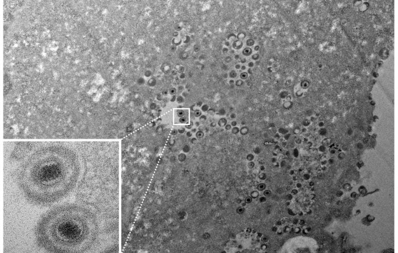
Stanford virologists are working with scientists at the new Stanford-SLAC Cryo-Electron Microscopy facility to take a new look at how herpesviruses infect cells.

Past Event

Presented by Wah Chiu. Cryogenic electron microscopy (cryo-EM) is a revolutionary technology for making 3D images of the inner workings of cells in much...

stillframe for public lecture
Video

Public lecture presented by Wah Chiu

stillframe for public lecture
Video
Public Lecture Poster
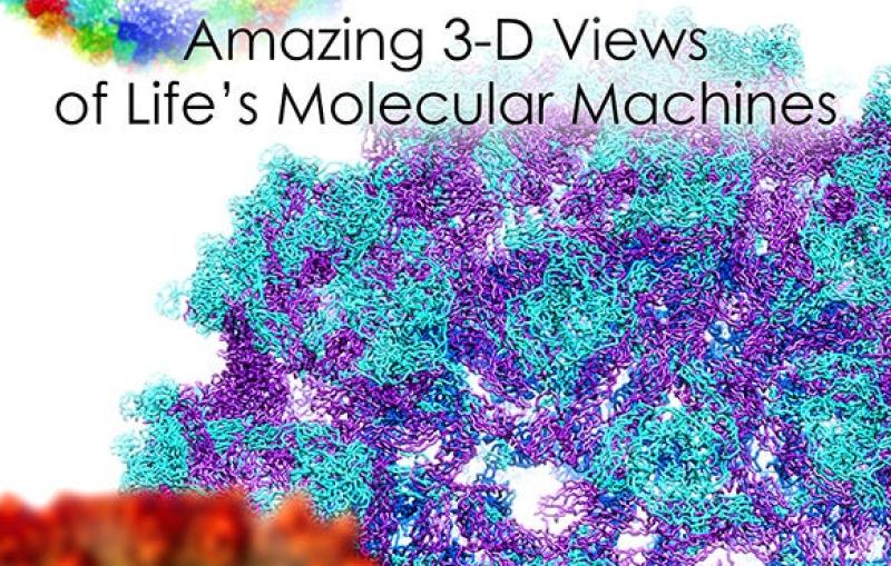
Public Lecture poster titled Cryo-EM: Amazing 3D Views of Life’s Molecular Machines
News Feature

This summer, five graduate students from the University of Puerto Rico had the opportunity to use SLAC’s world-class facilities to keep their studies on...

University of Puerto Rico Interns
News Feature

The National Institutes of Health center on the SLAC campus will make this revolutionary technology available to scientists nationwide and teach them how to...

Cryo-EM image of a proton pump involved in maintaining bone
Press Release

The new facility provides revolutionary tools for exploring tiny biological machines, from viral particles to the interior of the cell.

SLAC-Stanford Cryo-EM Facility
Press Release

Remarkable cryo-EM images that reveal details down to the individual atom will yield new insights into why high-energy batteries fail.

A lithium metal dendrite, taken with cryogenic electron microscopy or cryo-EM