Diamond-like Coating Improves Electron Microscope Images
Coating the surface of a material with a single layer of diamond-like crystals greatly improves images of it taken with an electron microscope, according to a study led by scientists at SLAC National Accelerator Laboratory and Stanford University.
By Mike Ross
Coating the surface of a material with a single layer of diamond-like crystals greatly improves images of it taken with an electron microscope, according to a study led by scientists at SLAC National Accelerator Laboratory and Stanford University.
In results published last month in Applied Physics Letters, the group reported a nearly three-fold improvement in the quality of photoelectron emission microscope (PEEM) images when they used the coating. PEEM images reveal important aspects of the sample’s surface structure, chemical bonding and magnetic properties.
The crystals, called diamondoids, are made of 10 or more carbon atoms arranged in the same way as a diamond. Known since the 1930s to exist in many oil and gas wells – where they are considered a nuisance – diamondoids are relatively cheap, readily available and the target of research by Stanford scientists who are exploring their properties and potential applications.
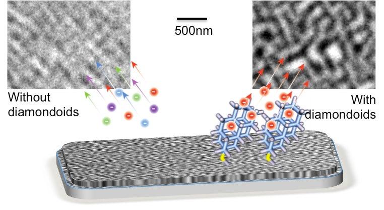
PEEM involves shining intense ultraviolet or X-ray light onto a sample and recording electrons emitted from just below its surface, which are used to form an image. However, the high X-ray energies often used in these studies trigger emissions of electrons with a wide range of energies – and this limits the resolution of the image. Although the image can be sharpened with complex million-dollar electronics, the Stanford/SLAC team thought a simple coating of diamondoids might work just as well – and it did.
“Earlier lab tests had shown that diamondoids should be excellent electron emitters,” said Nicholas Melosh, an associate professor of materials science and engineering at Stanford, and a researcher with the Stanford Institute for Materials and Energy Sciences (SIMES), a joint SLAC/Stanford institute. “This is the first published paper demonstrating that diamondoids can directly improve PEEM imaging.”
The tests were conducted at Lawrence Berkeley National Laboratory’s Advanced Light Source. Adding a single layer of diamondoid crystals to the surface of a cobalt-platinum magnetic alloy improved the resolution of the microscope image – the size of the smallest perceivable detail – from 25 nanometers, or billionths of a meter, to 10 nanometers. The diamondoids capture electrons emitted from the sample and re-emit them within a very narrow energy range, which can then be focused precisely.
“Our study exemplifies how the unique properties of a nanoparticle can help solve a problem not suited to conventional engineering solutions,” said SLAC microscopist Hendrik Ohldag. “With 10-nanometer resolution, we can start to identify and analyze the structures of domain walls – the critical transition regions between magnetic orientations in ferromagnetic or ferroelectric materials. It also enables us to significantly reduce the exposure time without sacrificing resolution, which is important when looking at organic materials, such as polymers, which degrade quickly in the X-ray beam.”
Future research aims to make the diamondoid coating more durable and find ways to use it on more materials, including non-metals. Ultimately, diamondoids could also improve the resolution of scanning electron microscopes, electron-beam writing and nanolithography tools. (However, recent price declines in flat-panel displays have reduced the likelihood that diamondoids would be used in those products.)
Stanford graduate student Hitoshi Ishiwata, who is investigating a range of novel diamondoid properties for his Ph.D. thesis, was lead author of the report. In addition to SLAC and Stanford scientists, the team included researchers from Berkeley Lab, Hitachi Global Storage Technologies (San Jose, Calif.), Swiss Federal Institute of Technology (Zurich) and Justus-Liebig University (Giessen, Germany).