Long-sought Discovery Fills in Missing Details of Cell 'Switchboard'
SLAC's X-ray Laser Lends New Insight into Key Target for Drug Development
Menlo Park, Calif. — A biomedical breakthrough, published today in the journal Nature, reveals never-before-seen details of the human body’s cellular switchboard that regulates sensory and hormonal responses. The work is based on an X-ray laser experiment at the Department of Energy’s SLAC National Accelerator Laboratory.
The much-anticipated discovery, a decade in the making, could have broad impacts on development of more highly targeted and effective drugs with fewer side effects to treat conditions including high blood pressure, diabetes, depression and even some types of cancer.
CXI | Long-sought Discovery Fills in Missing Details of Cell ‘Switchboard’
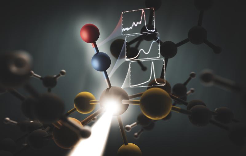
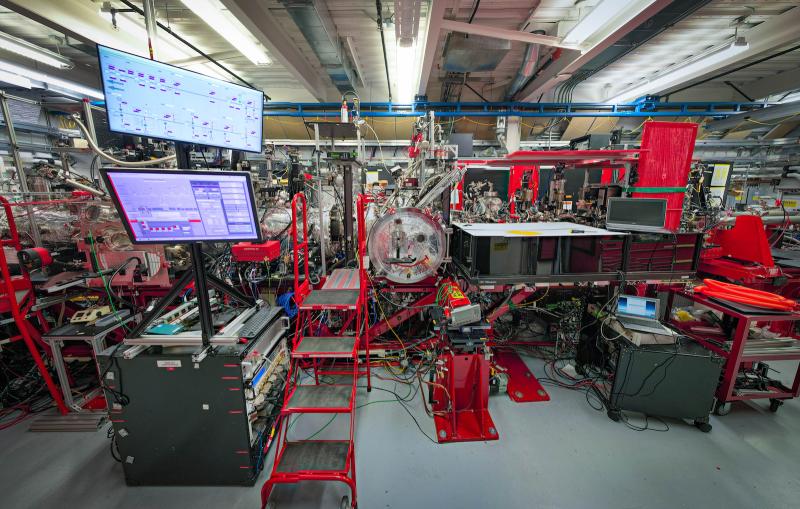
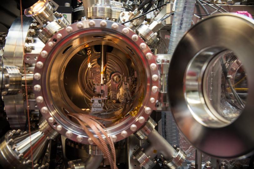
The study has been hailed by researchers familiar with the work as one of the most important scientific results to date using SLAC’s Linac Coherent Light Source (LCLS), a DOE Office of Science User Facility that is one of the brightest sources of X-rays on the planet. The LCLS X-rays are a billion times brighter than those from synchrotrons and produce higher-resolution images while allowing scientists to use smaller samples.
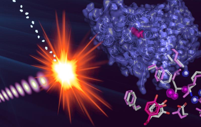
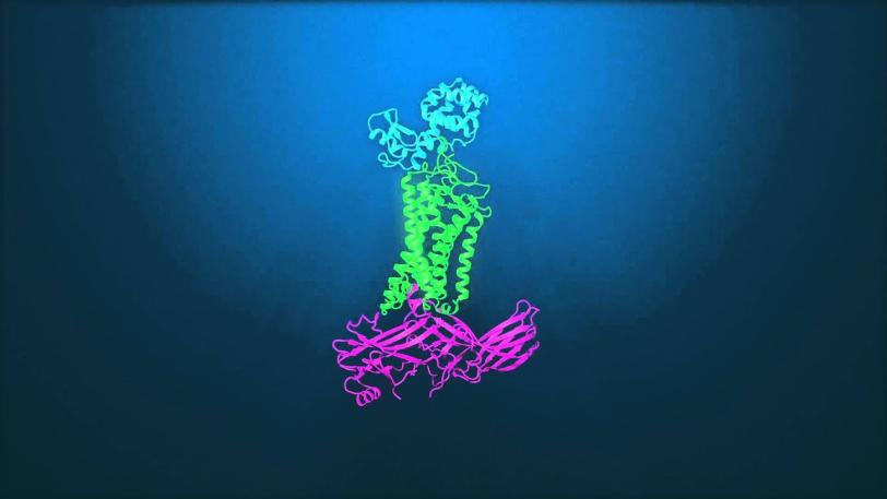
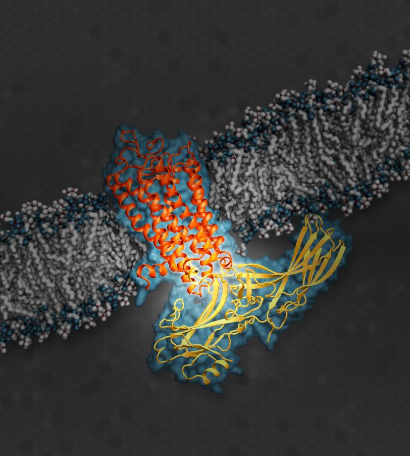
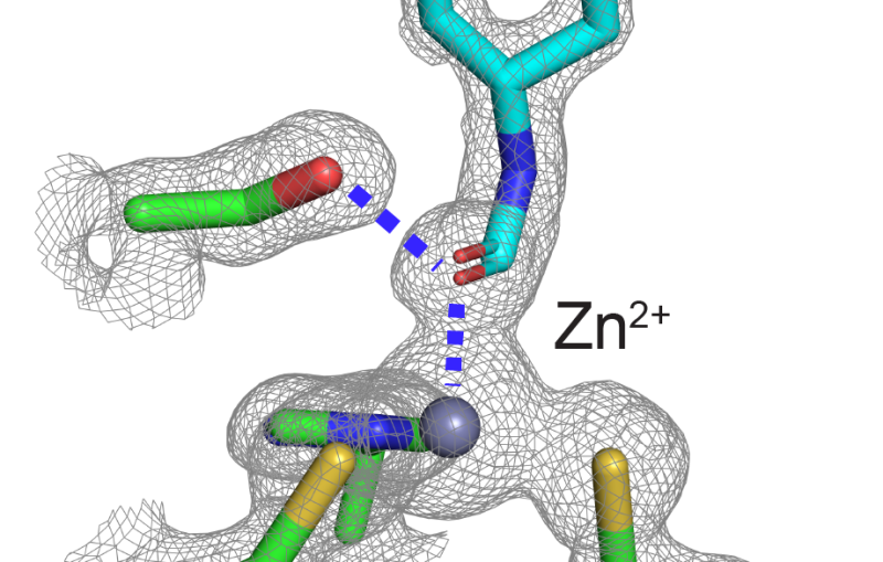
These ultrabright X-rays enabled the research team to complete the first 3-D atomic-scale map of a key signaling protein called arrestin while it was docked with a cell receptor involved in vision. The receptor is a well-studied example from a family of hundreds of G protein-coupled receptors, or GPCRs, which are targeted by about 40 percent of drugs on the market. Its structure while coupled with arrestin provides new insight into the on/off signaling pathways of GPCRs.
The research, led by scientists at the Van Andel Research Institute in Michigan in collaboration with dozens of other scientists from around the globe, represents a major milestone in GPCR structural studies, said Dr. Jeffrey L. Benovic, a biochemistry and molecular biology professor at Thomas Jefferson University in Philadelphia who specializes in such research but was not a part of this study.
“This work has tremendous therapeutic implications,” Benovic said. “The study is a critical first step and provides key insight into the structural interactions in these protein complexes.”
Decoding the Body's Cellular 'Switchboard'
Arrestins and another class of specialized signaling proteins called G proteins take turns docking with GPCRs. Both play critical roles in the body’s communications “switchboard,” sending signals that the receptors translate into cell instructions. These instructions are responsible for a range of physiological functions.
Until now, only a G protein had been seen joined to a receptor at this scale, one of the discoveries recognized with the 2012 Nobel Prize in Chemistry. Before the study at SLAC, little was known about how arrestins – which serve a critical role as the “off” switch in cell signaling, opposite the “on” switch of G proteins – dock with GPCRs, and how this differs from G protein docking. The latest research helps scientists understand how a docked arrestin can block a G protein from docking at the same time, and vice versa.
Many of the available drugs that activate or deactivate GPCRs block both G proteins and arrestins from docking.
“The new paradigm in drug discovery is that you want to find this selective pathway – how to activate either the arrestin pathway or the G-protein pathway but not both -- for a better effect,” said Eric Xu, a scientist at the Van Andel Research Institute in Michigan who led the experiment. The study notes that a wide range of drugs would likely be more effective and have fewer side effects with this selective activation.
X-ray Laser Best Tool for Tiny Samples
Xu said he first learned about the benefits of using SLAC’s X-ray laser for protein studies in 2012. The microscopic arrestin-GPCR crystals, which his team had painstakingly produced over years, proved too difficult to study at even the most advanced type of synchrotron, a more conventional X-ray source.
In the LCLS experiments, Xu’s team used samples of a form of human rhodopsin – a GPCR found in the retina whose dysfunction can cause night blindness – fused to a type of mouse arrestin that is nearly identical to human arrestin. Measuring just thousandths of a millimeter, the crystals – which had been formed in a toothpaste-like solution – were oozed into the X-ray pulses at LCLS, producing patterns that when combined and analyzed allowed researchers to reconstruct a complete 3-D map of the protein complex
“While this particular sample serves a specific function in the body, people may start to use this research as a model for how GPCRs, in general, can interact with signaling proteins,” Xu said. His team had been working toward this result since 2005.
SLAC Director Chi-Chang Kao said of the research milestone, “This important work is a prime example of how SLAC’s unique combination of cutting-edge scientific capabilities, including its expertise in X-ray science and structural biology, are playing key roles in high-impact scientific discoveries.”
Data Analysis Helps Fill in Missing Piece
Qingping Xu, a scientist in the Joint Center for Structural Genomics at SLAC’s Stanford Synchrotron Radiation Lightsource who helped to solve the 3-D structure, said it took many hours of computer modeling and data analysis to help understand and refine its details.
“This structure is especially important because it fills in a missing piece about protein-binding pathways for GPCRs,” he said. Even so, he noted that much work remains in determining the unique structures and docking mechanisms across the whole spectrum of GPCRs and associated signaling proteins.
Eric Xu said his group hopes to conduct follow-up studies at LCLS with samples of GPCRs bound to different types of signaling proteins.
In addition to scientists from SLAC, including LCLS and SSRL’s Joint Center for Structural Genomics, and the Van Andel Research Institute, the study also included researchers from: Arizona State University, University of Southern California, DESY lab’s Center for Free Electron Laser Science in Germany, National University of Singapore, New York Structural Biology Center, The Scripps Research Institute, University of California, Los Angeles, University of Toronto, Vanderbilt University, Beijing Computational Science Research Center in China, the University of Wisconsin-Milwaukee, Chinese Academy of Sciences, Paul Scherrer Institute in Switzerland, Trinity College in Ireland, University of Chicago, University of Konstanz in Germany, Chinese Academy of Sciences, Center for Ultrafast Imaging in Germany, and University of Toronto.
SLAC is a multi-program laboratory exploring frontier questions in photon science, astrophysics, particle physics and accelerator research. Located in Menlo Park, California, SLAC is operated by Stanford University for the U.S. Department of Energy Office of Science.
SLAC National Accelerator Laboratory is supported by the Office of Science of the U.S. Department of Energy. The Office of Science is the single largest supporter of basic research in the physical sciences in the United States, and is working to address some of the most pressing challenges of our time. For more information, please visit science.energy.gov.
Citation: Y. Kang, et al., Nature, 22 July 2015 (10.1038/nature14656)
Press Office Contact: Glenn Roberts, glennr@slac.stanford.edu, (650) 926-4496
Related stories
A newly published protein structure helps explain how some anti-cancer immunotherapy treatments work
Stanford-SLAC study shows how modifying enzymes’ electric fields boosts their speed
A newly published protein structure helps explain how some anti-cancer immunotherapy treatments work
Stanford-SLAC study shows how modifying enzymes’ electric fields boosts their speed