SLAC X-ray Laser Brings Key Cell Structures into Focus
Experiment is Important Step Toward Atomic-Scale Imaging of Intact Biological Particles
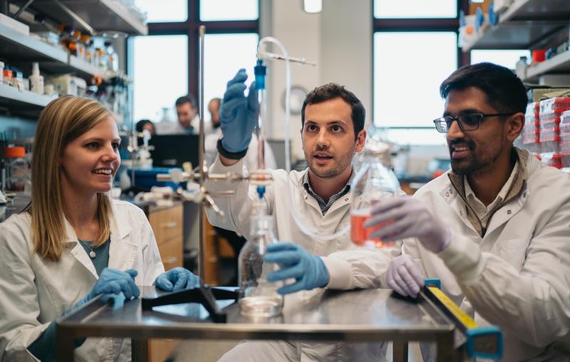
Scientists have made high-resolution X-ray laser images of an intact cellular structure much faster and more efficiently than ever possible before. The results are an important step toward atomic-scale imaging of intact biological particles, including viruses and bacteria.
The technique was demonstrated at the Linac Coherent Light Source (LCLS) at the Department of Energy’s SLAC National Accelerator Laboratory and reported in the Nov. 17 issue of Nature Photonics.
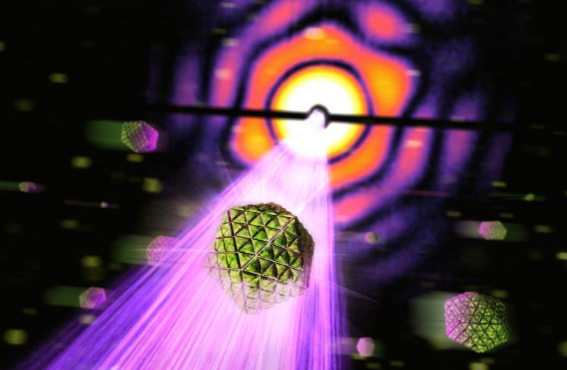
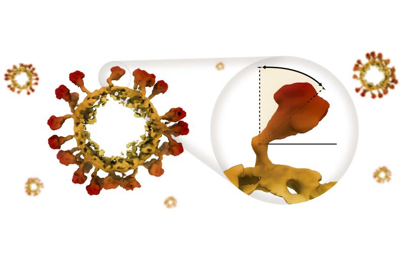
An international team of scientists used LCLS, a DOE Office of Science User Facility, to produce tens of thousands of high-resolution images of the tiniest individual biological particles yet studied there – 20-sided structures called carboxysomes that are found in some bacterial cells and play a major role in Earth’s life-sustaining carbon cycle. Although these structures have been studied for years, much remains unknown about their inner workings.
New Views of Bacteria’s Carbon-conversion Engines
"In an experiment lasting only 12 minutes, we collected many high-quality images using a very small amount of sample," said Janos Hajdu, a professor of biophysics at Uppsala University in Sweden, which led the research. "In the future you could conceivably get results within minutes that used to take five days for certain types of samples. We showed that with the right type of instruments and detectors, you can increase the throughput of X-ray lasers by a huge amount.”
The results bolster the aims of the LCLS Single-Particle Imaging initiative, formally launched at SLAC in October in cooperation with the international scientific community, said Christoph Bostedt, a SLAC senior staff scientist who leads the initiative with staff scientist Andy Aquila. The initiative is working toward atomic-scale imaging for many types of biological samples by identifying and addressing technical challenges at LCLS.
“As this work demonstrates, we are already achieving imaging of single particles today, and we are working to improve upon these capabilities,” Aquila said.
Bostedt added, “We will tackle these hurdles by working with the community. LCLS managers have committed to support this effort with dedicated experimental time.”
Biology ‘On the Fly’
Carboxysomes are microscopic workhorses inside cyanobacteria, which carry out photosynthesis. They pull in carbon dioxide from their surroundings and use an enzyme to convert it to forms that other living things can use. An estimated 40 percent of the organic carbon on Earth was made in these cell structures. "A better understanding of the structure and function of these cell organelles could benefit carbon-cycle research," said Dirk Hasse of Uppsala University, one of the paper’s lead authors.
Because they vary in size from about 90 to 140 nanometers, or billionths of a meter, in diameter, it is difficult to crystallize these tiny structures for conventional X-ray studies of their structure.
A 2011 study by members of the same international team had studied samples of a giant virus, called Mimivirus, using a similar experimental technique that jets samples in a finely focused aerosol spray into the path of the X-ray pulses.
In the latest study they were able to achieve better resolution, showing features as small as 18 nanometers.
The scientists said a new design for the “aerosol injector” that shot the samples in a thin stream toward the X-ray pulses was among the key improvements that allowed them to achieve higher-resolution images. They also improved data-analysis tools that automatically sorted the 70,000 images they collected and assembled them into distinct groups. These advances lay the foundation for accurate, high-throughput structure determination for individual biological samples and other types of samples.
The resolution achieved in this first experiment was not high enough to reveal the interior of the carboxysomes, though the team said that upgrades to higher intensity and a narrower focus of LCLS X-rays could allow far more detailed explorations of many biological samples in this size range, including small viruses. “We haven’t yet reached the limits,” said Filipe Maia of Uppsala University, who participated in the study.
Pushing the Limits
Max Hantke of Uppsala University, a lead author of the study, said the ultrashort pulses of X-ray lasers offer important advantages over instruments like electron microscopes that are used to study biological samples.
For example, the X-ray laser can be used to track ultrafast processes, such as how biological samples respond to pulses of light over time. “These types of experiments are not possible with other methods,” Hantke said.
In addition to scientists from Uppsala University and SLAC, other researchers who participated in the study were from Lawrence Berkeley National Laboratory; Kansas State University; the National University of Singapore; the University of Melbourne in Australia; University of Rome in Italy; and DESY laboratory, PNSensor, Max Planck Institute for Extraterrestrial Physics and European XFEL, all in Germany.
This work was supported by the Swedish Research Council, the Knut and Alice Wallenberg Foundation, the European Research Council, the Röntgen-Ångström Cluster, and Stiftelsen Olle Engkvist Byggmästare. The CAMP instrument used in the experiment was funded by the Max Planck Society.
Citation: Max F. Hantke, Dirk Hasse, et al., Nature Photonics, 17 November 2014; (10.1038/nphoton.2014.270).
For questions or comments, contact the SLAC Office of Communications at communications@slac.stanford.edu.
SLAC is a multi-program laboratory exploring frontier questions in photon science, astrophysics, particle physics and accelerator research. Located in Menlo Park, Calif., SLAC is operated by Stanford University for the U.S. Department of Energy's Office of Science.
SLAC National Accelerator Laboratory is supported by the Office of Science of the U.S. Department of Energy. The Office of Science is the single largest supporter of basic research in the physical sciences in the United States, and is working to address some of the most pressing challenges of our time. For more information, please visit science.energy.gov.
Related stories
A newly published protein structure helps explain how some anti-cancer immunotherapy treatments work
Stanford-SLAC study shows how modifying enzymes’ electric fields boosts their speed
A newly published protein structure helps explain how some anti-cancer immunotherapy treatments work
Stanford-SLAC study shows how modifying enzymes’ electric fields boosts their speed