From Parchment to People: SLAC Invention Measures Stroke Damage in the Brain
A technique SLAC scientists invented for scanning ancient manuscripts is now being used to probe the human brain, in research that could lead to new medical imaging methods and better treatments for stroke and other brain conditions.
By Glennda Chui
A technique SLAC scientists invented for scanning ancient manuscripts is now being used to probe the human brain, in research that could lead to new medical imaging methods and better treatments for stroke and other brain conditions.
The studies, taking place at SLAC’s Stanford Synchrotron Radiation Lightsource, are led by cell biologist Helen Nichol, of the University of Saskatchewan, with $2.5 million in funding from the Canadian government and the Heart and Stroke Foundation of Canada.
Her team, which includes a Stanford neurosurgeon and stem cell expert, other medical doctors and experts in stroke research and medical imaging, reflects the broad ambitions of this research: to give doctors a better understanding of what they’re seeing in MRI scans of stroke patients; to improve diagnosis and guide treatments; and maybe even to develop new ways to peer inside the living brain. What all these goals have in common is that they depend on the ability to track movements and deposits of tiny traces of metal in human tissue. That’s a job the SSRL technique, known as rapid-scanning XRF, is exquisitely suited to do.
At a synchrotron equipped with this technology, “you can see a large sample of brain, and you have the high resolution the technique offers to actually zoom in on your single cells,” said Dr. Raphael Guzman, a pediatric neurosurgeon and stem cell expert at Stanford University Medical Center who is leading part of the study.
Regular XRF, or X-ray fluorescence imaging, uses the SSRL’s powerful X-ray beam to knock electrons out of the inner shells of atoms in a sample. As more electrons fall in to fill the gaps, they give off light – fluoresce – and the color of that light reveals which chemical elements are present.
In the mid-2000s, SLAC scientists had a chance to use this technique to examine a priceless manuscript – the Archimedes palimpsest, a 10th century parchment containing copies of works by the ancient Greek mathematician that had been erased by monks and recycled as a prayer book. But they soon realized that to examine something this big in a reasonable amount of time, they would have to make the scanning go much faster.
Led by physicist Uwe Bergmann, they developed a way to move the beam continually over the sample, rather than imaging one spot at a time. This allowed them to proceed 300 times faster – a scan that used to take 12 days could now be done in an hour – and opened up the possibility of examining much bigger samples, from art objects and cultural artifacts to fossils of early birds. In 2006, Bergmann and an international team of researchers used rapid-scanning XRF to reveal the words of Archimedes, including passages that had been lost for centuries, beneath the prayer writings on the old parchment.
When she read about this research, Nichol said, “It just grabbed me. I thought, if he could map something as big as a sheet of paper, we could map a brain.”
She and her colleagues began using rapid-scanning XRF at the SSRL to look at metals in the preserved brains of people who had died with Alzheimer’s disease, Parkinson’s disease or multiple sclerosis. The healthy brain needs metals like iron, zinc, manganese and copper to work properly, and some studies had indicated that in people with neurodegenerative diseases, the normal distribution of these metals was out of whack. But did these changes cause the disease, or were they a result of it? And were they consistent enough to offer a tool for diagnosis?
To the team’s disappointment, scans of brain slices from eight people with Parkinson’s disease found no clear pattern – nothing that could help doctors diagnose their brain conditions or understand how they came about. “What we found is that the changes you see in Parkinson’s and Alzheimer’s are sort of variations on normal,” Nichol said.
She decided that beam time on the SSRL was better spent studying a disorder that caused clear, obvious damage in the brain. Stroke fit the bill.
When a stroke blocks the flow of blood to the brain, it produces striking lesions, almost like bruises, caused by bleeding and tissue death. Blood contains iron, which is part of the hemoglobin that carries oxygen in red blood cells. When bleeding occurs, the iron leaks out in a form that can damage surrounding cells, so the body quickly tucks the iron away in various chemical compounds for safe storage.
Standard MRI scans can image and identify those iron compounds and show doctors where bleeding has taken place. But they may not catch the very smallest bleeds, Nichol said, or identify other elements that may be disrupted during a stroke.
That’s where RS-XRF comes in. As the first practical tool for imaging a number of different metals in all of their chemical forms at the same time – and over a large section of the brain – it “opens up a lot of doors to things you can’t see with medical imaging,” Nichol said. It also can tell one form of iron from another; the spectrum of iron in hemoglobin will look different than free-floating iron or the iron compounds produced by bleeding, for instance.
The idea behind the study is that iron in its varied forms can be used as a marker to reveal changes in molecules and cells that follow a stroke, evaluate stroke damage and follow the migration of stem cells that are injected into patients in experimental stroke treatments. The scientists will also look at sulfur compounds that are thought to play a role in protecting the brain from damage, and evaluate the effects of the few stroke treatments available, such as chilling the brain, on the distribution of iron and sulfur.
Members of the team come to the SSRL about three weeks per year to scan brain tissue from rats, including some that have been bred to make them unusually susceptible to stroke, as well as human brain tissue from the National Institutes of Health brain bank. Additional experiments are being done at the Canadian Light Source at the University of Saskatchewan.
While they are not putting live patients in a synchrotron, the scientists hope their findings will someday result in the ability to scan live patients with methods that are much more sensitive to damage from tiny strokes that now go unnoticed.
In the meantime, by comparing the results of MRI scans with RS-XRF scans of the same tissue, the researchers hope to improve doctors’ ability to interpret what they see in MRI images.
“It’s one thing for me to show people a pretty picture that shows there may be iron present and another thing to validate that – to quantify how much is there, how much has spread throughout the brain,” said Mark Haacke of Wayne State University in Detroit, a specialist in medical imaging who is playing a leading role in the study. “XRF allows us to create a scale for measuring iron directly from an MR image. Knowing those details may help us understand why people suffer differently – why some people recover from stroke and some people don’t.”
Stanford’s Guzman has been labeling neural stem cells with nanoparticles of iron oxide, injecting them into the bloodstream and using RS-XRF to track them as they move into the brain.
“It has been found that you can take neural stem cells, transplant them into the brain and they will integrate – form connections and secrete proteins that will help the brain to heal,” he said. “A particularity of these cells is that once they reach the brain they migrate. They get attracted to the lesion and they actually move over a period of several weeks toward the lesion.”
By comparing brain images taken with MRI and rapid-scanning XRF, he said, he hopes to find a formula for translating the amount of the iron tagging compound in the brain tissue into the number of stem cells it represents. Down the road, he’d like to identify the receptors on the insides of blood vessels that grab the stem cells and facilitate their journeys into the brain.
The results “will have much broader applications than just stroke and the brain,” Guzman said. “We’re talking brain now, but there’s cardiac, there’s liver, there are many areas of injury” that may be treatable with stem cells. Learning how to track and image those stem cells, he said, will help in developing those therapies, too.
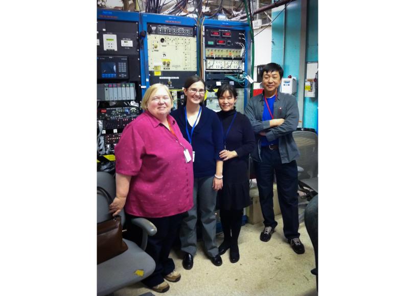
(Photo courtesy Raphael Guzman)
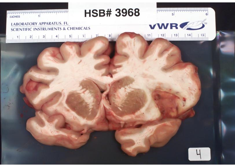
(Photo courtesy Helen Nichol)