Tools of the Trade: New X-ray Microscopy Technique Makes Faster, Sharper Images
A new x-ray microscopy technique that allows scientists to make images 60 times faster than before has been developed by an Australian research team including Garth Williams, who is now an instrument scientist at SLAC’s Linac Coherent Light Source.
By Mike Ross
A new x-ray microscopy technique that allows scientists to make images 60 times faster than before has been developed by an Australian research team including Garth Williams, who is now an instrument scientist at SLAC’s Linac Coherent Light Source. The method, called polychromatic coherent diffractive imaging, or polyCDI, will also enable sharper and more accurate images of floppy biological molecules and fast-moving materials structures.
“One of the longstanding problems arising from using coherent diffractive imaging to study the structure of samples at storage rings has been stability, especially with regard to 3-D structures due to the relatively long exposure times required, sometimes up to 10 seconds,” Williams said. “Sub-second exposure times will reduce this concern. This new technique occupies an important niche and will become widely available, initially at synchrotron light sources.”
To generate an x-ray image from CDI data, the sample is illuminated with a single wavelength of light, or as close to that ideal as possible. The x-rays interact with the atoms in the sample and diffract, producing a continuous pattern that can be used to reveal the sample shape and internal structure. The wavelength of the X-rays determines the angle of diffraction, so when many wavelengths are present in the illuminating beam the diffraction pattern becomes blurred.
Unfortunately, the most common sources of bright X-rays – synchrotrons – produce a somewhat broad range of wavelengths. Scientists working at synchrotrons use monochromators to block unwanted wavelengths, so only a narrow range of wavelengths illuminates the samples. While this does produce high-quality images, it also greatly reduces the intensity of the light hitting the sample and increases exposure times. This inevitably blurs images of soft, biological and rapidly changing samples.
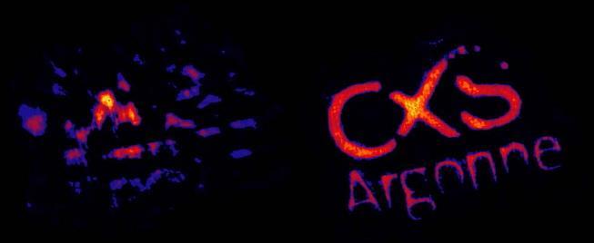
The Aussies came up with a mathematical solution. Rather than filtering the light with a monochromator, they let a greater fraction of the light hit the sample and used an intricate new type of analysis to calculate how each of its many wavelengths was diffracted. Combining all of those contributions in the correct ratio allows them to determine the structure of the molecule or material from the pattern of fuzzy spots. Using all the available light cuts exposure time dramatically and results in a much sharper image.
The initial demonstration was conducted at the Advanced Photon Source at Argonne National Laboratory in Illinois. A report describing polyCDI, along with its first images, was published in the June 26 online edition of Nature Photonics. The project was conducted by scientists from the Australian Research Council’s Center of Excellence for Coherent X-ray Science, which is headed by Keith Nugent of the University of Melbourne. Williams was a postdoctoral fellow in Nugent’s research group when this research began. He and his colleagues helped develop the original concept for polyCDI, design the experimental geometry and collect early experimental data.
Scientists doing tomography and X-ray fluctuation microscopy can also benefit from the shorter exposure times, said Brian Abbey, a postdoctoral fellow in Nugent’s group who is the paper’s first author and leader of this new research: “It could also make table-top X-ray sources practical for CDI, enabling nanoscale biological X-ray imaging experiments in a typical laboratory setting.”
Williams added that polyCDI could ultimately improve the quality of some images at free-electron laser X-ray sources, such as the Linac Coherent Light Source, because even their highly coherent light pulses contain more than one wavelength of light.