Past Event · Public lecture

Cryo-EM: Amazing 3D Views of Life’s Molecular Machines

Presented by Wah Chiu

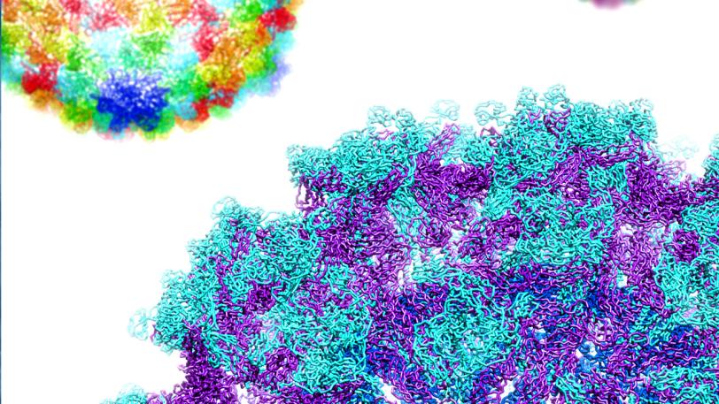
Cryogenic electron microscopy (cryo-EM) is a revolutionary technology for making 3D images of the inner workings of cells in much higher resolution than ever possible before. Under development for four decades, it’s seen such rapid progress over the past few years that three of its developers were awarded a Nobel prize in 2017. “Cryo” refers to the fact that samples are flash-frozen before being “photographed” from multiple angles with focused electron beams. The reconstructed images show how atoms are arranged within molecular machines inside our cells – from the proton channels that help keep cells healthy by controlling their acidity to the molecular gadgets viruses use to maintain their ability to infect. This lecture presents newly obtained images that show a wide variety of these tiny engines in action, and describe cryo-EM’s potential for improving human health.


About Wah Chiu

Wah Chiu received his PhD in biophysics from the University of California,  Berkeley in 1975, where he participated in the early days of cryo-electron microscope imaging of biological molecules. After appointments at Lawrence Berkeley National Laboratory and the University of Arizona, he moved to Baylor College of Medicine in Houston, where he founded a major center for the study of protein structure and biological imaging. In 2017, he returned to California as a Stanford professor with a joint appointment in bioengineering, microbiology and immunology and in photon science at SLAC, where he is the director of the lab’s new national user facility for cryo-electron microscopy. Chiu is a member of the National Academy of Sciences.

Past Event · Public lecture

Cryo-EM: Amazing 3D Views of Life’s Molecular Machines

Presented by Wah Chiu

Public Lectures
stillframe for public lecture
Video
Date
Tuesday, September 25, 2018
12:30–1:30 p.m. PDT