See content related to electron diffraction and electron microscopy techniques here below.
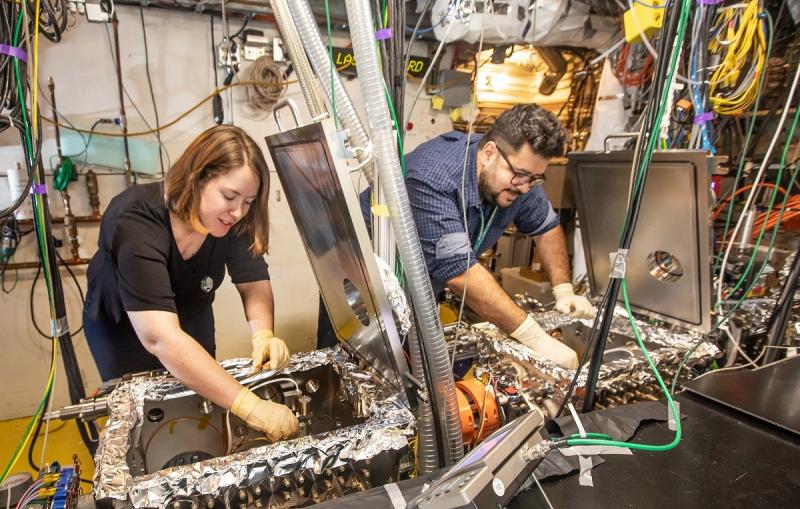
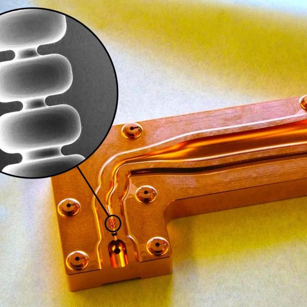
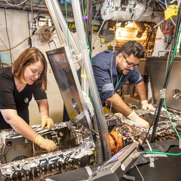
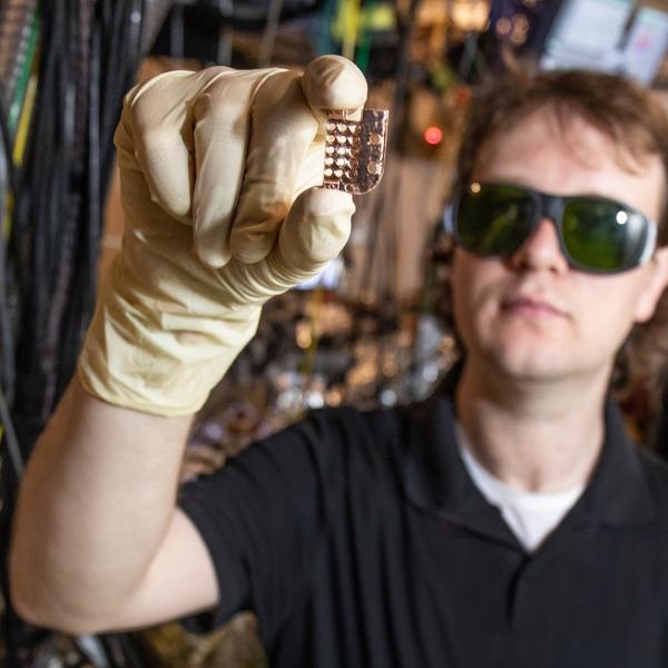
SLAC’s Emma Snively and Mohamed Othman at the lab’s high-speed “electron camera,” an instrument for ultrafast electron diffraction (MeV-UED).
(Jacqueline Orrell/SLAC National Accelerator Laboratory)
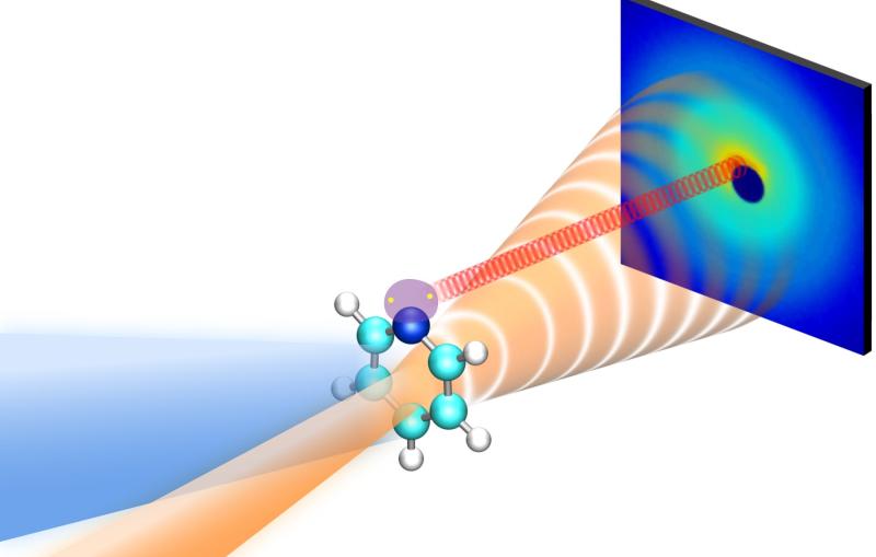
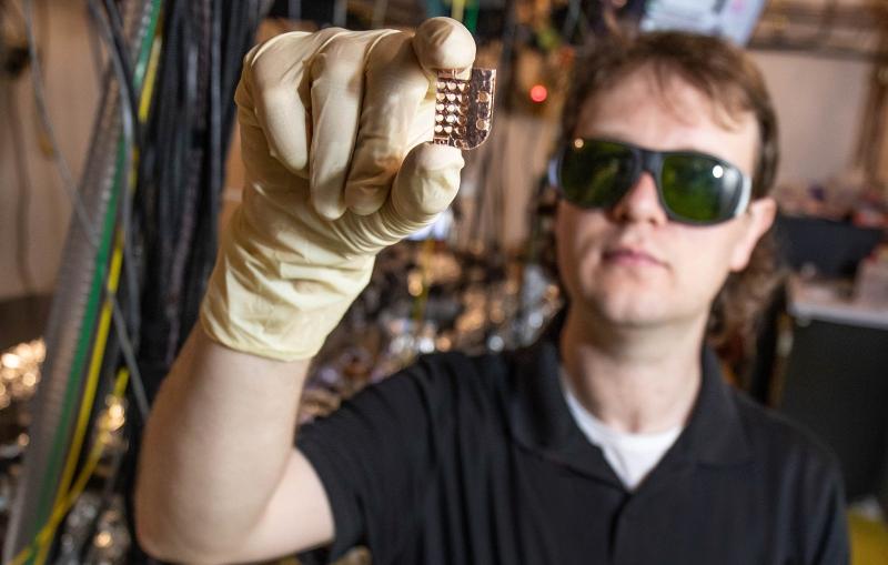
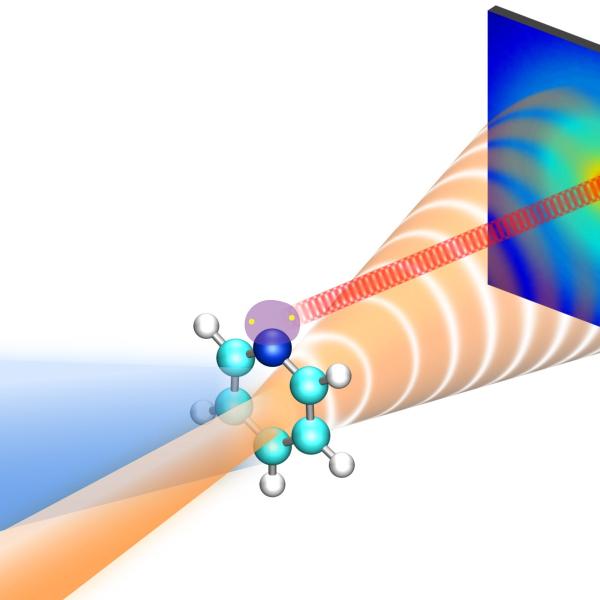
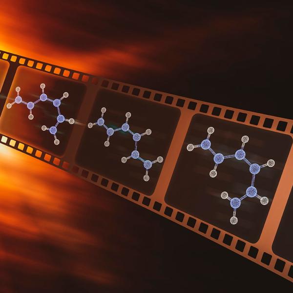
This new technology could enable future insights into chemical and biological processes that occur in solution, such as vision, catalysis and photosynthesis.
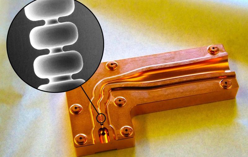
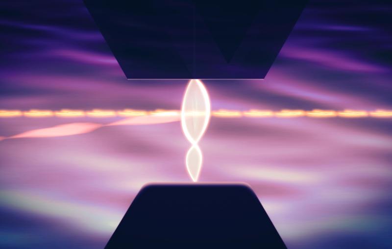
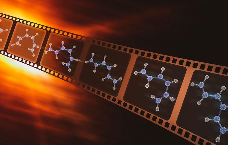
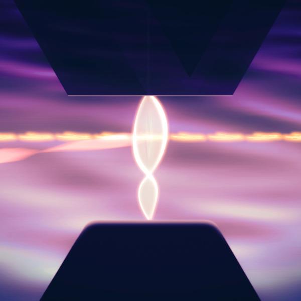
Researchers have squeezed a high-energy electron beam into tight bundles using terahertz radiation, a promising advance in watching the ultrafast world of atoms unfold.
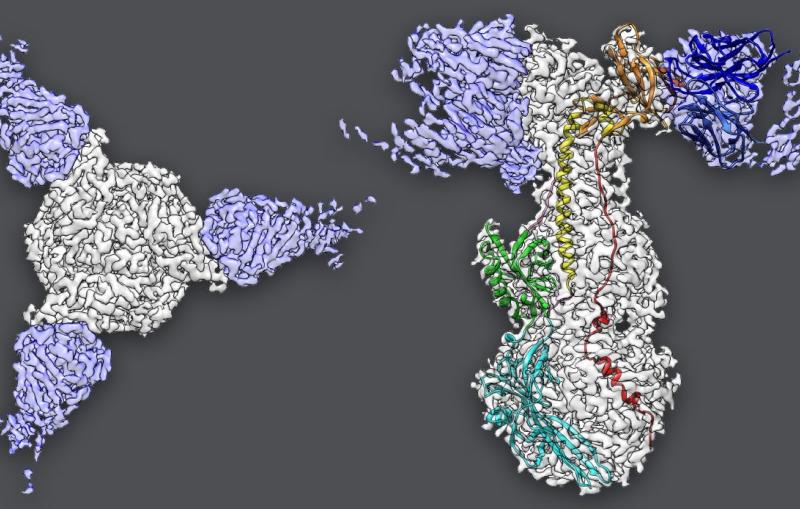
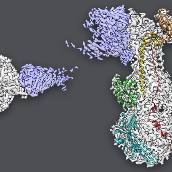
Cryogenic electron microscopy can in principle make out individual atoms in a molecule, but distinguishing the crisp from the blurry parts of an image...
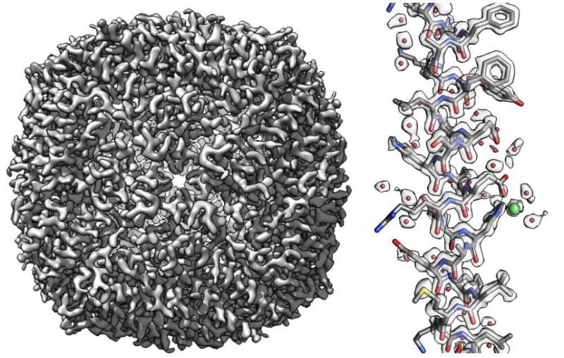
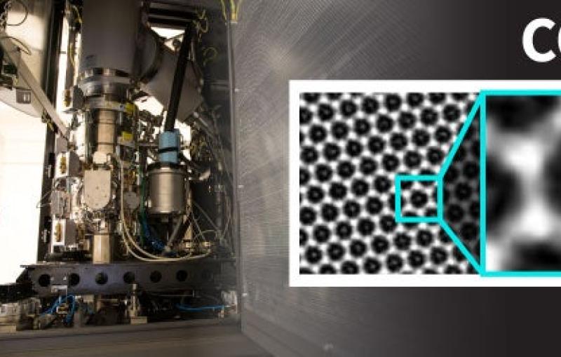
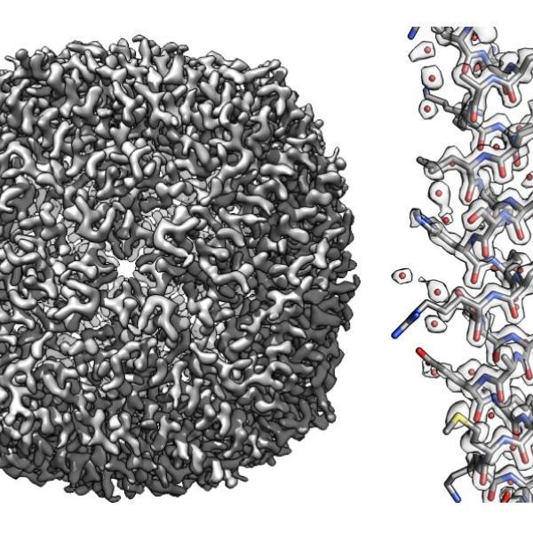
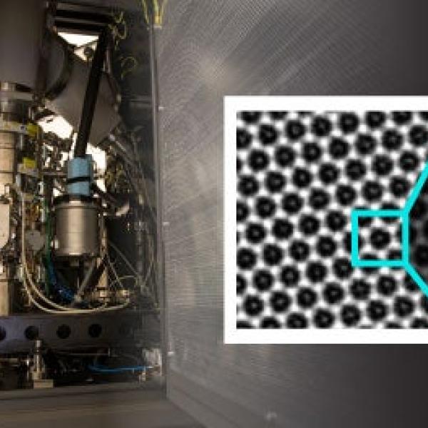
A new twist on cryo-EM imaging reveals what’s going on inside MOFs, highly porous nanoparticles with big potential for storing fuel, separating gases and...
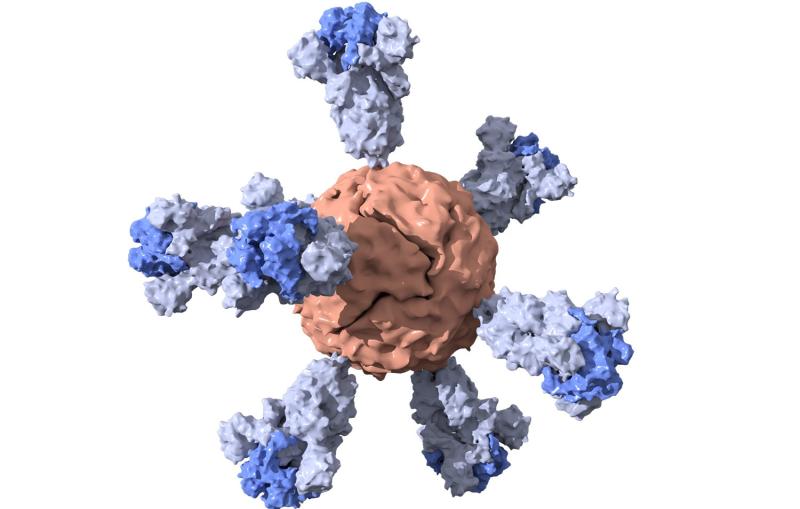
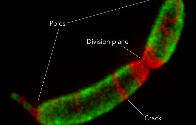
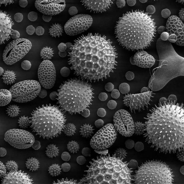
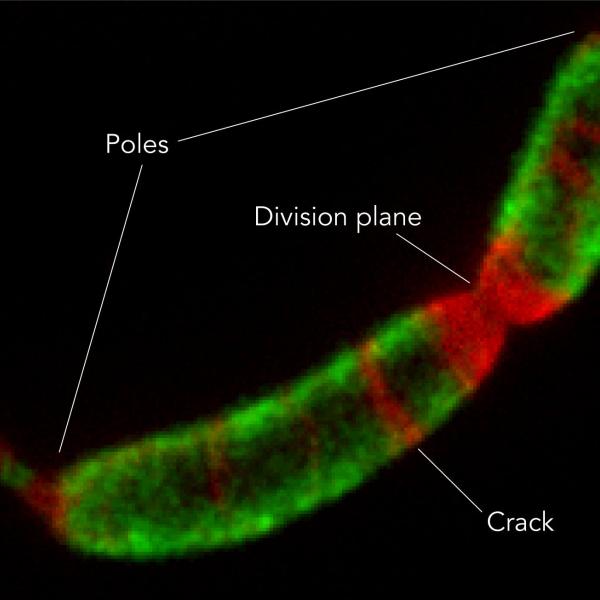
A close-up look at how microbes build their crystalline shells has implications for understanding how cell structures form, preventing disease and developing nanotechnology.
This new technology could enable future insights into chemical and biological processes that occur in solution, such as vision, catalysis and photosynthesis.
Researchers have squeezed a high-energy electron beam into tight bundles using terahertz radiation, a promising advance in watching the ultrafast world of atoms unfold.
Cryogenic electron microscopy can in principle make out individual atoms in a molecule, but distinguishing the crisp from the blurry parts of an image can be a challenge. A new mathematical method may help.
A new twist on cryo-EM imaging reveals what’s going on inside MOFs, highly porous nanoparticles with big potential for storing fuel, separating gases and removing carbon dioxide from the atmosphere.
A close-up look at how microbes build their crystalline shells has implications for understanding how cell structures form, preventing disease and developing nanotechnology.