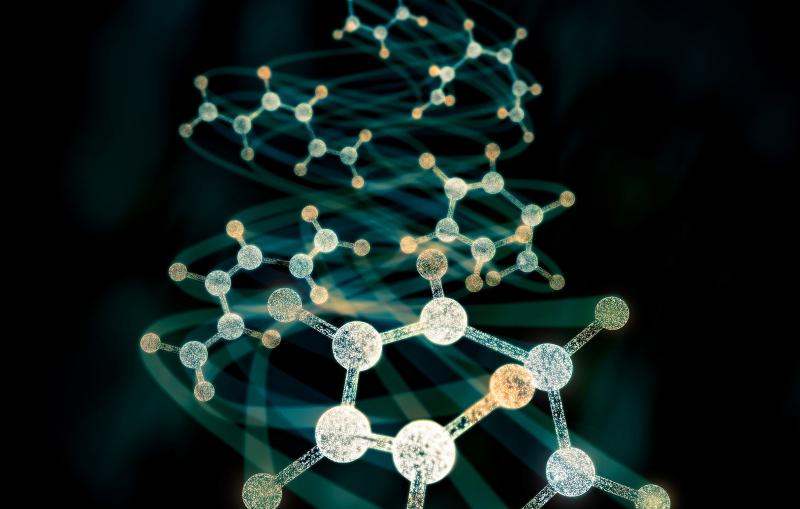
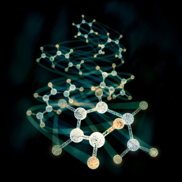
Tiny microbes and molecular machines have an outsized impact on human health, and they play key roles in the vast global cycles that shape climate and make carbon and nitrogen available to all living things.
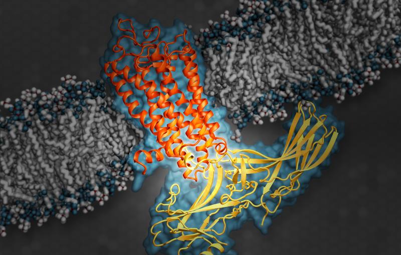
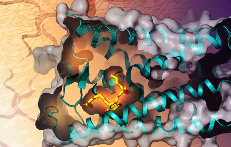
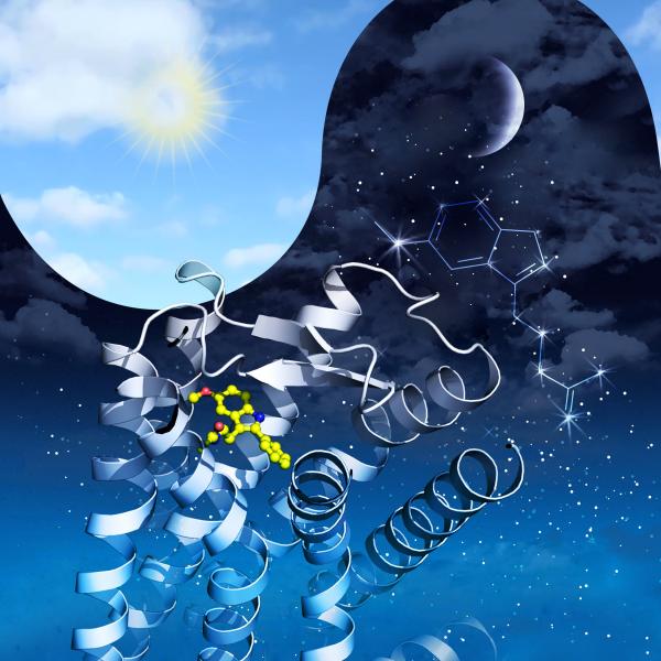
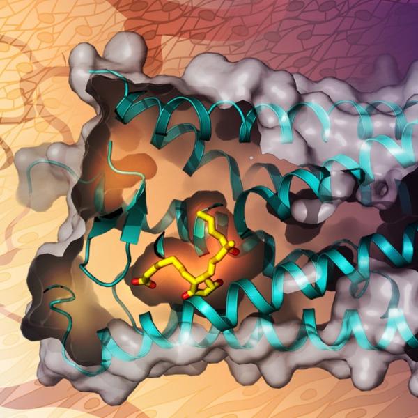
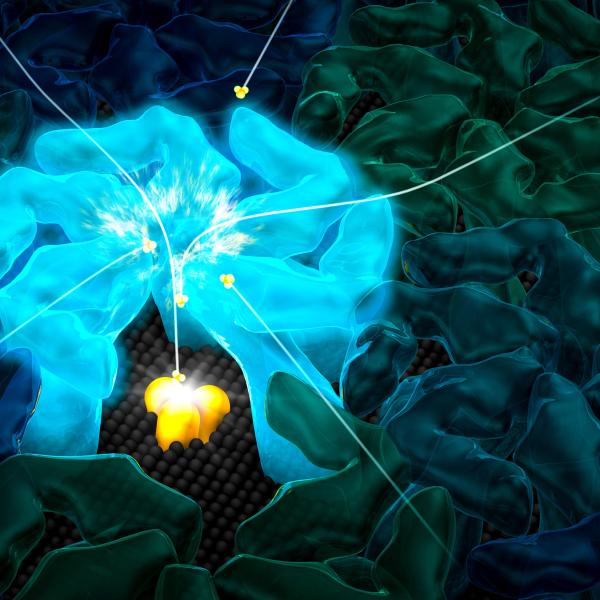
This illustration shows arrestin (yellow), an important type of signaling protein, while docked with rhodopsin (orange), a G protein-coupled receptor.
(Greg Stewart/SLAC National Accelerator Laboratory)
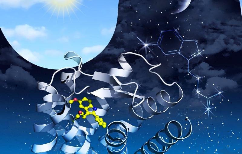
A better understanding of how these receptors work could enable scientists to design better therapeutics for sleep disorders, cancer and Type 2 diabetes.
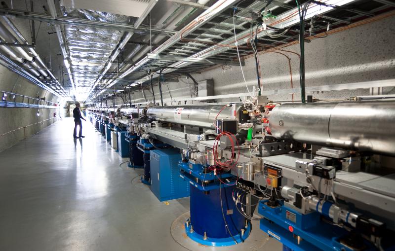
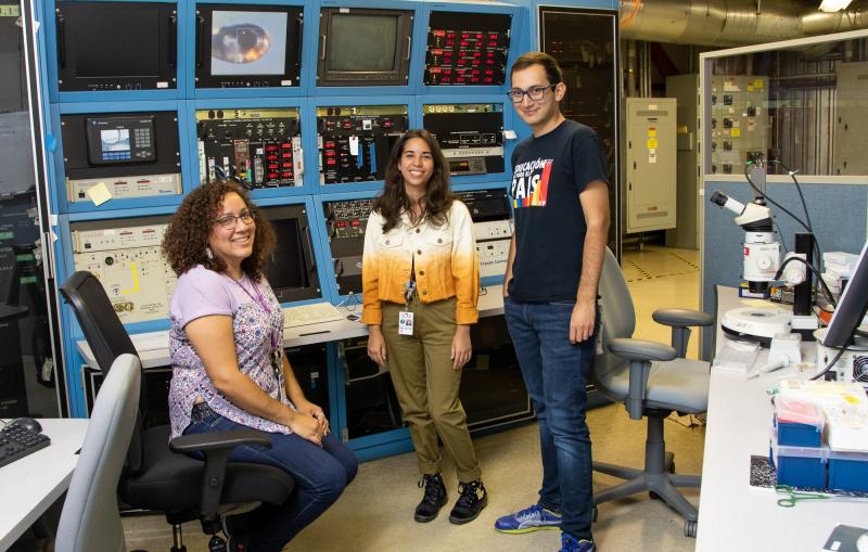
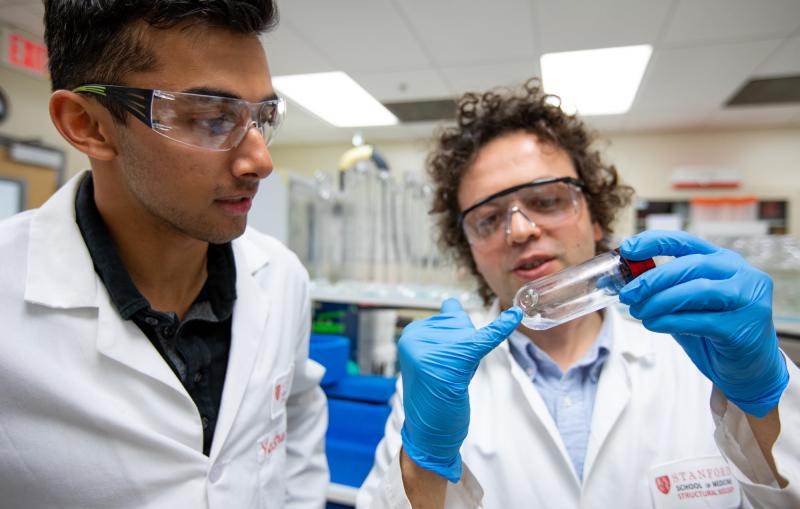
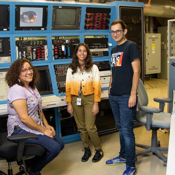
This summer, five graduate students from the University of Puerto Rico had the opportunity to use SLAC’s world-class facilities to keep their studies on...
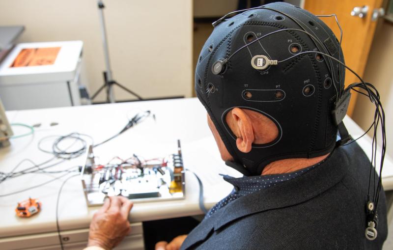
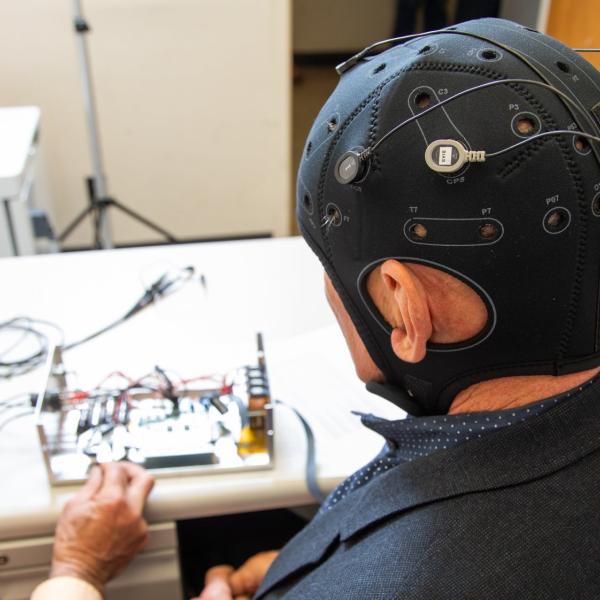
SLAC and Stanford researchers are developing a device that combines electrical brain stimulation with EEG recording, opening potential new paths for treating neurological disorders.
A better understanding of how these receptors work could enable scientists to design better therapeutics for sleep disorders, cancer and Type 2 diabetes.
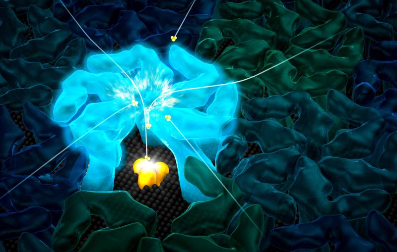
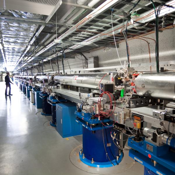
In a major step forward, SLAC’s X-ray laser captures all four stable states of the process that produces the oxygen we breathe, as well as fleeting steps in between. The work opens doors to understanding the past and creating a...
This summer, five graduate students from the University of Puerto Rico had the opportunity to use SLAC’s world-class facilities to keep their studies on track.
SLAC and Stanford researchers are developing a device that combines electrical brain stimulation with EEG recording, opening potential new paths for treating neurological disorders.
The X-ray laser movie shows what happens when light hits retinal, a key part of vision in animals and photosynthesis in microbes. The action takes place in a trillionth of an eye blink.
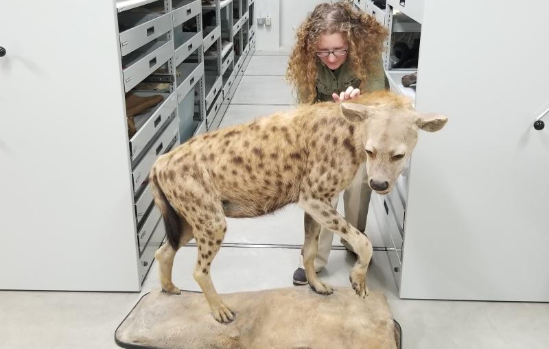
Tiny pores in the shells of archaea microbes attract ammonium ions that are their sole source of energy, allowing them to thrive where this food is so scarce that scientists can’t even detect it.